RCM1 is an easy to use, sensitive, high resolution and affordable confocal imaging system:
An ideal solution for small labs with limited budget, but demanding tasks, particularly when high sensitivity and resolution are desired from the imaging system.
A confocal microscope that works as a camera, no need for an instruction manual.
RCM1 is extremely easy to use: no hardware control or software processing needed, and the images are always RAW.
RCM1 can be delivered as a total microscope system with a selection of microscopes (Nikon, Olympus, Leica or Zeiss), a selection of cameras (Hamamatsu, PCO, Andor, Photometrics) and laser solutions (Omicron, Toptica).
If you already have a microscope in the lab, RCM1 is an upgrade to an existing wide-field fluorescence system – RCM1 can easily be added to the existing wide-field fluorescence microscope system to improve its resolution.
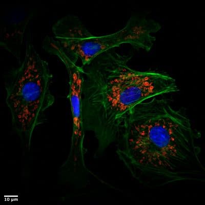
Standard mode – high resolution RCM image. FOV 80 x 80 micron
Increased FOV – standard confocal resolution. FOV 160 x 160 micron
Who should use RCM1 and why?
Obtain super-resolution confocal images with standard dyes and without complicated training. It’s a fruitful solution for individual research labs to easily convert any existing widefield fluorescence microscope into a super-resolution and highly sensitive confocal system. Record living cells and organoids in 4D super-resolution using very low laser power. Don’t worry about phototoxicity or photobleaching. RCM1 provides optimized conditions for live-cell imaging and facilitates posterior analysis with sharp and high contrast raw images.
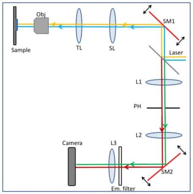
RCM Working Principle
The RCM technique extends standard confocal microscopy with a re-scanning unit, improving lateral resolution by √2 and reducing signal to noise ratio.
Re-scan Confocal Microscopy (RCM) is a new super-resolution technique based on standard confocal microscopy extended with an optical (re-scanning) unit that projects the image directly on a CCD-camera. This new microscope has improved lateral resolution (170 nm at 488 nm excitation), and strongly improved sensitivity, while maintaining the sectioning capability of a standard confocal microscope. It is particularly useful for biological applications where the combination of high-resolution and high-sensitivity is required (but not very high imaging speed).
The excitation lasers (blue and yellow lines) are directed via a dichroic mirror towards the first scanning unit SM1. As in a standard confocal microscope, the scanning unit scans the laser light in the sample and de-scans the emission light, directing it at the pinhole PH (green and red lines). After the pinhole, a second re-scan unit SM2 directs the light onto a camera chip.[/caption]
During scanning, re-scan mirrors (SM2) move faster than the first scan mirrors (SM1). This magnifies the image on the camera chip compared to the sample, and eventually results in the higher resolution of the image. The resolution of the system is improved with the re-scan step by a factor of √2 (i.e. 1.41 times), compared to Abbe’s resolution limit by changing the angular amplitude of the re-scanner (SM2). Reduction of pinhole is no longer necessary to increase resolution. Closing down the pinhole only limits the amount of light passing through and decreases the signal to noise ratio due weaker signal. Since the re-scan is a purely optical method with no further image processing required, there is cost in time while improving the resolution. By using a sensitive camera as detector, the signal-to-noise ratio of the RCM is 4 times higher than in standard confocal microscopy.
To fully understand the principle of rescanning, resolution improvement and the optical layout of the RCM, please watch the video below that explains the components and the light path of the RCM (animation credits to StudioFlip). Additional technical details and test images can be found in De Luca et al (2013).
The Re-scan Confocal Microscopy (RCM) module can be used to turn any fluorescent microscope into a confocal microscope. For an upgrade, laser(s) and a camera are needed.
Check magnificent Turnkey examples from our customers for your inspiration:
Let us know which equipment you have available, and we will make a custom tailored upgrade solution for you.
Axial Resolution
500 nm (350 after deconvolution)
Lateral Resolution
170 nm (120 nm after deconvolution)
Quantum Efficiency
70-95% (camera dependent)
Scan Speed
1 FPS @ 512 x 512 pixels
FOV
130 x 130 μm (60x, super resolution
Detector
Camera
Optimized for
100x, 60x (high NA)
Scanner
Analog (open loop)
Wavelength
VIS-NIR
Software
Micromanager, Nis Elements, Volocity
Integration
API
PSF for deconvolution
Microvolution, SVI Huygens
Bypass Mode
Yes
Check out the More Information tab for comparison of RCM with Widefield, Confocal and other similar systems!
Confocal imaging
RCM has improved lateral resolution and the same axial resolution compared to conventional confocal microscope.
Ratio-imaging and multi-color applications
RCM can work in multi-colour mode for different colour combinations and ratio-imaging (applications like FRET, FRAP, pH, and Ca2+ imaging).
Live cell imaging
RCM has a very high signal-to-noise ratio and quantum efficiency (80-95%, depending on the camera), which means no need for high laser power.
HO1N1 cells stained with Bacmam 2.0 Mito-GFP (green), ER-tracker (blue) and SiR-DNA (red). One 3-color image was taken every 10 seconds for 1.5 hours, giving a total of 1000 images. Because the RCM is very sensitive laser power can be kept very low (few microwatts), enabling long-term imaging. The native lateral resolution of the RCM is 170 nm. After deconvolution by SVI Huygens the resolution is improved to 120 nm! Movie taken by Jeroen Kole (Confocal.nl), sample courtesy Dandan Ma (ACTA, Amsterdam), equipment provided by Marko Popovic (Nikon Center of Excellence, Amsterdam University Medical centers, VUmc, Amsterdam.)
HO1N1 cells expressing Mitochondria-RFP through the Bacmam expression system. One image was taken every 10 seconds for 61 hours (giving a total of almost 22,000 images!). Laser power was measured to be 1 microwatt at the sample plane. Movie taken by Jeroen Kole (Confocal.nl), sample courtesy Dandan Ma (ACTA, Amsterdam), equipment provided by Marko Popovic (Nikon Center of Excellence, Amsterdam University Medical centers, VUmc, Amsterdam.)
The first video shows the RAW data from RCM, the entire 61 hour time lapse.
The second video shows the benefits of deconvolution. We used SVI Huygens CMLE deconvolution to improve the resolution to 120 nm and have an even better signal-to-noise ratio.
Images taken with RCM1
References
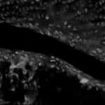
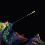
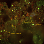
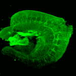
[1] Fibroblast stained with tubulin – Alexa 790. Imaged with 25x air objective. Sample courtesy Gertrude Bunt, Universitätsmedizin Göttingen . Image by Desiree Salas Pastene (Confocal.nl). [2] Ancinus (hollow secretory organoid) formed by growing mammary epithelial cells (MCF-10A cells) in 3D culture (embedded in Matrigel) for 12 days. Cells are stained for mtHSP70 (mitochondrial marker, Red), Paxillin (Green) and DNA (DAPI, Blue). Image courtesy of Dr R. Pedley , A. Gilmore’s group, Wellcome Trust Centre for Cell-Matrix Research. [3]Unstained mouse knee, 50um-thick: 3D sections with 405nm excitation (top view). Endogenous fluorescence from chondrocytes in articular cartilage. Sample courtesy of Zhiyi Liu, Li Zeng and Irene Georgakoudi, Department of Biomedical Engineering, Tufts University, Medford, USA and Sackler School of Graduate Biomedical Sciences, Tufts University, Boston, USA. [4] Triple labeling of actin filaments, as described by Resnik et al in https://doi.org/10.1007/s00418-019-01806-3. Sample courtesy of Natasha Resnik, Institute of Cell Biology, Faculty of Medicine, University of Ljubljana. [5] Cultured cells with labeling of chromosomes (blue) and exosomes (red; small structures visible in the image on the right). Specimens by Gert van Capellen (Erasmus MC), images by Erik Manders. [6] Autofluorescence in a soil sample. Courtesy Elly Morriën, UVA, Amsterdam. [7] Hair Follicle Stem Cells. Sample courtesy of Yulia Shwartz, Ya-Chieh Hsu lab, SCRB, Harvard University. [8] Primary culture of hippocampal neurons from rat embryos. Labelling: Alpha-tubulin (Abberior STAR 635P) in magenta and mitochondria (Abberior STAR 580) in yellow. Samples prepared by Laurent Ladépêche and Jesús Planagumà, labelling by Maria Marsal. Images taken and processed by Marina Cunquero, ICFO – The Institute of Photonic Sciences.
Deconvolution
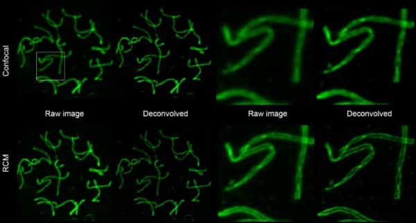
Representative nuclear spread from fixed mouse spermatocytes, immunostained for SYCP3 a component of the synaptonemal complex (Alexa 488-labelling) imaged with RCM using without re-scanning (confocal) and with re-scanning (RCM). Left: full spread. Right: individual chromosomes. Sample courtesy of A. Agostinho – Advanced Light Microscopy Facility, Science for Life Laboratory
C. Höög – Department of Cell and Molecular Biology, Karolinska Institutet. Imaging and deconvolution by Erik Manders (Confocal.nl). For deconvolution SVI Huygens software was used.
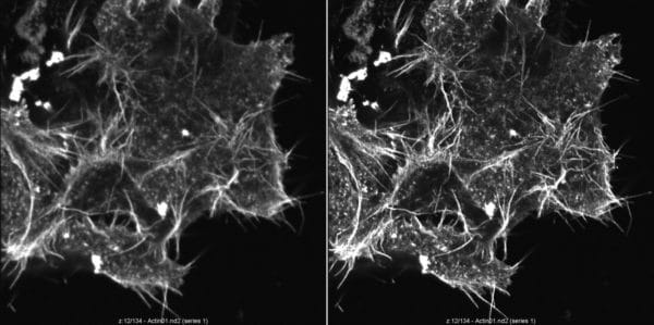
N1E-115 neuroblastoma cells, made during HOLM 2017 by the students attending one of the workshops. On the left, raw image from RCM, on the right the same image after the Microvolution deconvolution
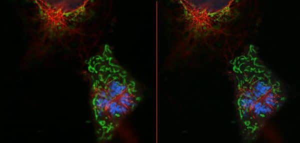
Mitotic cell – Cos7 cell line with antibody labeling of nucleus (blue), mitochondria (green), and tubulin (red). Samples prepared by Jana Doehner (University of Zurich). Images taken by Stan Hilt (Confocal.nl) during the BioImaging meeting Utrecht. Original RCM image (left), and the result of NIS Elements deconvolution (right).
3D Reconstructions
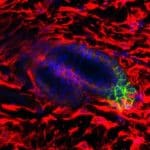
Hela cell in mitosis stained for DAPI (blue), Actin (green) and Tubulin (red). Sample courtesy of Nicolas Touret, Faculty of Biochemistry, University of Alberta
Doll S, Freitas FP, Shah R, Aldrovandi M, da Silva MC, Ingold I, Grocin AG, Xavier da Silva TN, Panzilius E, Scheel CH, Mourão A, Buday K, Sato M, Wanninger J, Vignane T, Mohana V, Rehberg M, Flatley A, Schepers A, Kurz A, White D, Sauer M, Sattler M, Tate EW, Schmitz W, Schulze A, O’Donnell V, Proneth B, Popowicz GM, Pratt DA, Angeli JPF, Conrad M. FSP1 is a glutathione-independent ferroptosis suppressor. Nature. 2019 Oct 21. doi: 10.1038/s41586-019-1707-0.
The RCM scanning confocal unit can fit to any inverted or upright microscope via C-mount adapter between the microscope and the camera. The laser light is fed into the RCM unit via a single mode FC/APC fiber connector. The RCM comes in two versions: RCM-VIS and RCM-NIR. Both can operate up to 4 colors, RCM-VIS is design to operate with visible light (350-700 nm), while RCM-NIR is designed to operate in the Near-Infrared spectrum (650-1050 nm). The RCM comes with a bypass module to operate the camera in widefield mode.
Microscopes
RCM can be mounted to any inverted or upright microscope. Upright microscope requires the Upright Microscope Mounting Kit. Motorised focussing unit (internal or external) is also needed in most applications. Currently, RCM has been tested and approved for the following microscopes: Nikon Ti & Ti2, Olympus IX, Zeiss Axio Observer, Leica DMi6000 & DMi8 and MiCube. Contact us if you have any question regarding microscope compatibility.
Please note that:
RCM only mounts correctly to the original microscope manufacturers C-mount
A 100/0 beam splitter is mandatory. 50/50, 70/30 and 80/20 beam splitters do not work with RCM, as they are not laser safe.
Objective lenses
In order to obtain the 170 nm resolution, the highest NA (>1.40) lenses available have to be used.
Cameras
Any microscope camera with a C-mount connector, external trigger, and similar specifications as the two first ones in the list below should work with the RCM. Please contact us, if you have a camera that you would like to test for the RCM compatibility.
We offer Hamamatsu Flash 4 V2 (LT) and V3, which is tested to work with RCM. Please let us know if you have a camera to verify the compatibilty
A physical pixel size of 6.5 micron or lower is ideal for 60-100x objective lenses, as this results in sufficiently small sampling. At 100x objective magnification and 6.5 micron pixel the sampling is 43 nm at the sample plane.
Beamsplitter
Hamamatsu W-View Gemini A12801-01
Cairn Multisplit V2, four-way emission splitter
Software
RCM drivers are available for LabView (free of charge), Micro-Manager / ImageJ (Fiji) (free of charge), and Nikon NIS Elements (at cost).
Lasers
We offer Oxxius laser combiner. The RCM laser coupling is compatible with single mode fibers with FC/APC connector and NA 0.11. Using other fiber connectors will degrade image quality.
Wavelengths: For RCM-Visible lasers with a wavelength from 405 upwards are suitable. RCM is designed as a 4 color system. We offer a flexible laser solution that can be equipped with 1 to 4 lasers. Laser lines can be upgraded at later stage as well.
As the RCM is sensitive, we only need low power lasers (max 25 mW). The advantage of the low power lasers is the significantly longer lifetime and lower costs.
Rescan Confocal Microscope Comparisons
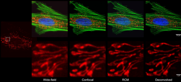
RCM vs Wide-field & Confocal
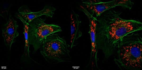
In order to demonstrate the image improvement by RCM, we have made images of the same cell from deer skin fibroblast sample using wide-field, conventional confocal microscope and RCM. The image taken by RCM we have further improved using deconvolution.
Indian Muntjac – Deer Skin Fibroblast cells. Staining: Blue: DAPI, Green: Phalloidin-Alexa488, Red: Mitotracker CMXRos
Top: image from full field of view; bottom: zoom in. Images by Jeroen Kole (Confocal.nl).
Method
Microscope
Objective
Detector
Pinhole
Wide-field
Zeiss Axiovert 200M
63x NA 1.4; oil
PCO sensicam (100.3nm/pixel)
Confocal
Nikon A1
60x NA 1.4; oil
GaAsP PMT
1 AU
RCM
RCM +Nikon TiE Eclipse
100x NA 1.45; oil
Hamamatsu Orca Flash 4.0
1 AU
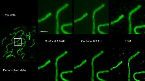
RCM vs PMT-based confocal system
The resolution and sensitivity of the raw RCM image is better, resulting in a better deconvolution result. The light intensity at the sample plane was measured at: 4.5 microwatt (1.0 AU), 12.5 microwatt (0.3 AU) and 3.0 microwatt (RCM).
Nuclear spread from fixed mouse spermatocytes, immunostained for SYCP3 a component of the synaptonemal complex (Alexa 488-labelling). Upper panel: images of a PMT based confocal with 1.0 AU pinhole, 0.3 AU pinhole and RCM image. Lower panel: same images deconvolved using Huygens Essential RCM module (SVI), using an experimental point spread function. Scalebar: 1 micrometer. Sample courtesy of A. Agostinho – Advanced Light Microscopy Facility, Science for Life Laboratory. Imaging and deconvolution performed by Jeroen Kole (Confocal.nl).
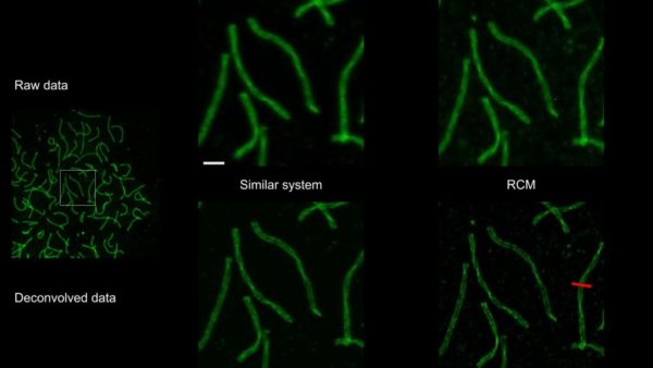
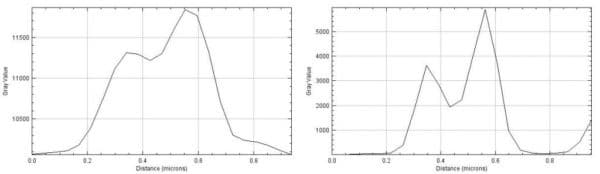
RCM vs similar system
We have compared the performance of RCM with that of similar image scanning system (based on technology described in DOI:10.1103/PhysRevLett.104.198101 ). After SVI deconvolution with a measured point spread function the resolution of the deconvolved RCM data is better as shown by the lineplots of the red line.
Nuclear spread from fixed mouse spermatocytes, immunostained for SYCP3 a component of the synaptonemal complex (Alexa 488-labelling). Upper panel: raw images obtained with a similar system* and RCM image. Lower panel: same images deconvolved using a measured point spread function. Scalebar: 1 micrometer. Sample courtesy of A. Agostinho – Advanced Light Microscopy Facility, Science for Life Laboratory. Imaging and deconvolution performed by Jeroen Kole (Confocal.nl).
RCM Large FOV vs RCM High resolution
RCM can be used in different imaging modes: Large Field of View allows maximizing the view, but at expense of resolution. In this mode, conventional confocal resolution is achieved. High Resolution, is the RCM mode providing improvement in resolution, but at the expense of the size of view.
Rescan confocal microscope is an easy to use, sensitive, high resolution and affordable confocal imaging system:
An ideal solution for small labs with limited budget, but demanding tasks, particularly when high sensitivity and resolution are desired from the imaging system,
A confocal microscope that works as a camera, no need for an instruction manual
RCM is extremely easy to use: no hardware control or software processing needed, and the images are always RAW.
We can deliver the Rescan Confocal Microscope as a total microscope system with a selection of microscopes ( Nikon, Olympus, Leica or Zeiss ), a selection of cameras ( Hamamatsu, PCO, Andor, Photometrics ) and laser solutions (Omicron, Toptica ).
In case you already have a microscope in the lab, Rescan Confocal Microscope is an upgrade to an existing wide-field fluorescence system – RCM can easily be added to the existing wide-field fluorescence microscope system to improve its resolution.
The new Rescan Confocal Microscope (1.1)
The new RCM (1.1) is based on RCM (1.0) and has all the same features. On top of these, the new RCM offers:
Bypass mode
Two times the field of view
A scanning speed up to 4 frames per second
Integration in SVI Huygens deconvolution software
RCM 1.1 is available in a VIS and NIR version.
Resolution: the standard resolution of the RCM is 170 nm at 488 nm wavelength. This is also called super-resolution. In contrast to other super-resolution systems, RCM offers the super-resolution of the live (raw) image without any processing. It is possible to improve the RCM resolution even further, to 120 nm using deconvolution.
Frame rate: the frame rate of the RCM is 1 fps at 512 x 512, which makes the acquisition time of a 3 colour image about 3 seconds. The frame rate of RCM 1.1 can be increased to 4 fps at 512 x 512, which makes the acquisition time of a 3 colour image less than 1 second. With the new RCM 1.1 applications that require a bit faster imaging are now possible.
Field of view: Rescan Confocal Microscope 1.0 has a Field of View (FOV) of 80 x 80 μm at 100x magnification, RCM 1.1 has the option to increase this FOV to 160 x 160 μm.
Standard mode – high resolution RCM image. FOV 80 x 80 micron
Increased FOV – standard confocal resolution. FOV 160 x 160 micron
Rescan Confocal Microscope Models
Working Principle
The Rescan Confocal Microscopy technique extends standard confocal microscopy with a re-scanning unit. It improves lateral resolution by √2 and reduces signal to noise ratio.
Rescan Confocal Microscope(RCM) is a new super-resolution technique based on standard confocal microscopy extended with an optical (re-scanning) unit that projects the image directly on a camera chip. This new microscope has improved lateral resolution (170 nm at 488 nm excitation), and strongly improved sensitivity, while maintaining the sectioning capability of a standard confocal microscope. It is particularly useful for biological applications that requires combination of high-resolution and high-sensitivity (but not very high imaging speed).
Diagram
The excitation lasers (blue and yellow lines) are directed via a dichroic mirror towards the first scanning unit SM1. As in a standard confocal microscope, the scanning unit scans the laser light in the sample and de-scans the emission light, directing it at the pinhole PH (green and red lines). After the pinhole, a second re-scan unit SM2 directs the light onto a camera chip.
During scanning, re-scan mirrors (SM2) move faster than the first scan mirrors (SM1). This magnifies the image on the camera chip compared to the sample, and eventually results in the higher resolution of the image. The re-scan step improves the resolution of the system by a factor of √2 (i.e. 1.41 times), compared to Abbe’s resolution limit by changing the angular amplitude of the re-scanner (SM2). Reduction of pinhole is no longer necessary to increase resolution. Closing down the pinhole only limits the amount of light passing through and decreases the signal to noise ratio due weaker signal. Since the re-scan is a purely optical method with no further image processing required. There is a cost in time for improving the resolution. By using a camera as a detector, the SNR of the RCM is 4 times higher than the standard confocal microscopy.
To fully understand the principle of rescanning, resolution improvement and the optical layout of the RCM, please watch the video below that explains the components and the light path of the RCM (animation credits to StudioFlip). Additional technical details and test images can be found in De Luca et al (2013).
")

 Description
 Specifications
 Applications
 Pictures
 Videos
 Downloads
 Publications
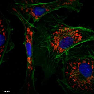


 Working Principle