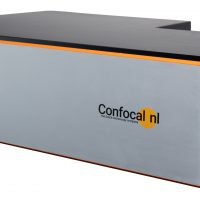
 Description
 Specifications
 Applications
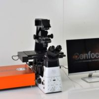
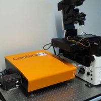
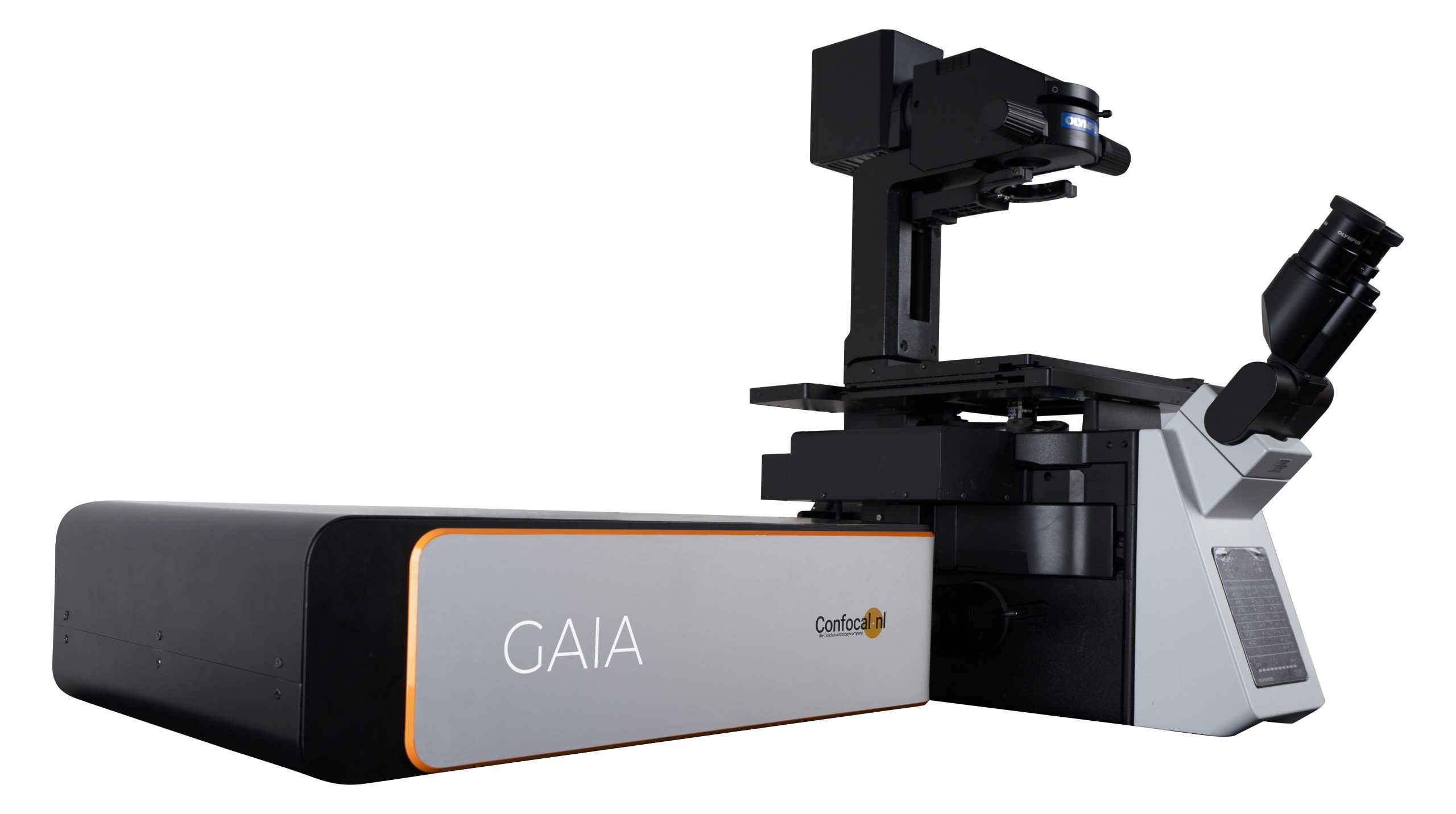
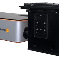
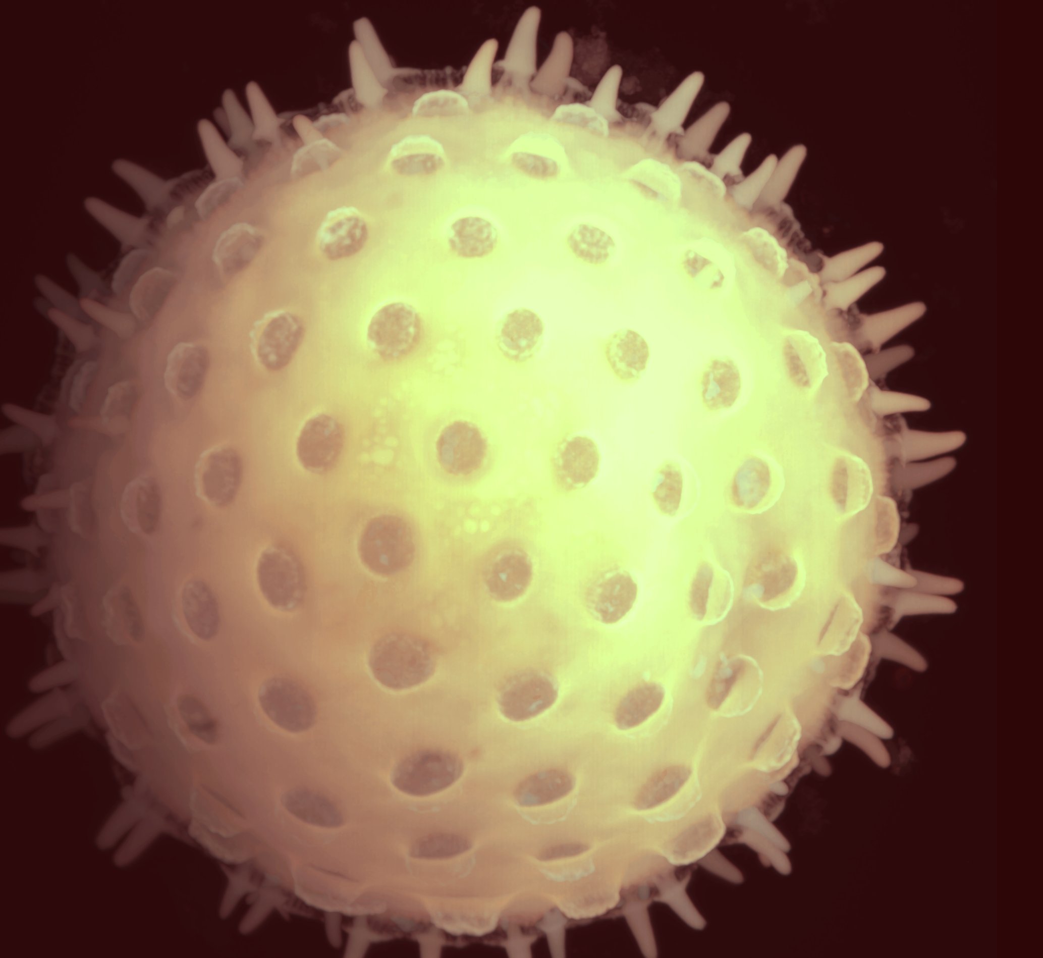
 Pictures
 Downloads
 Publications
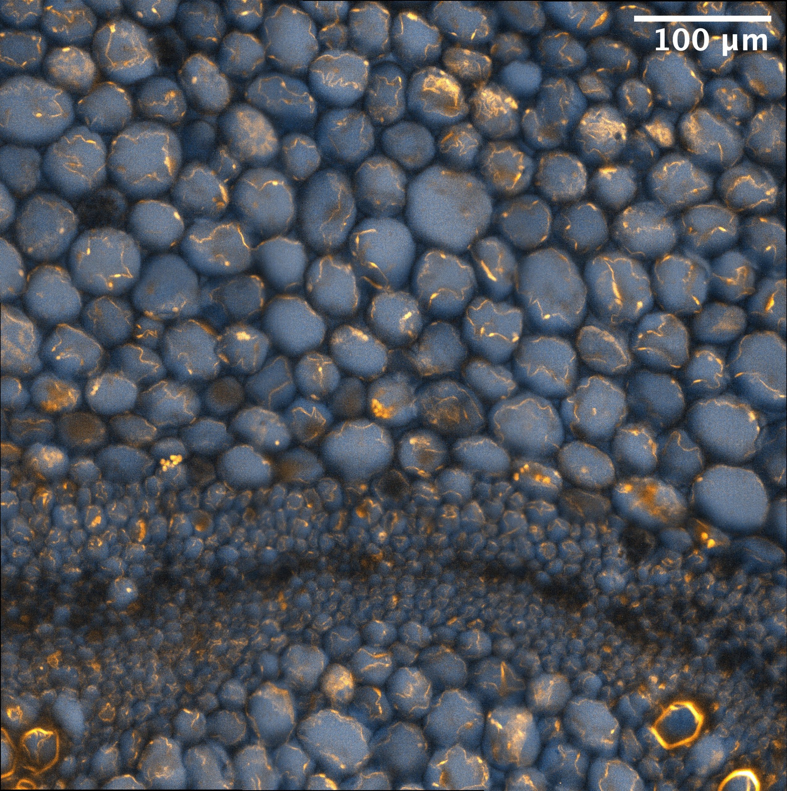
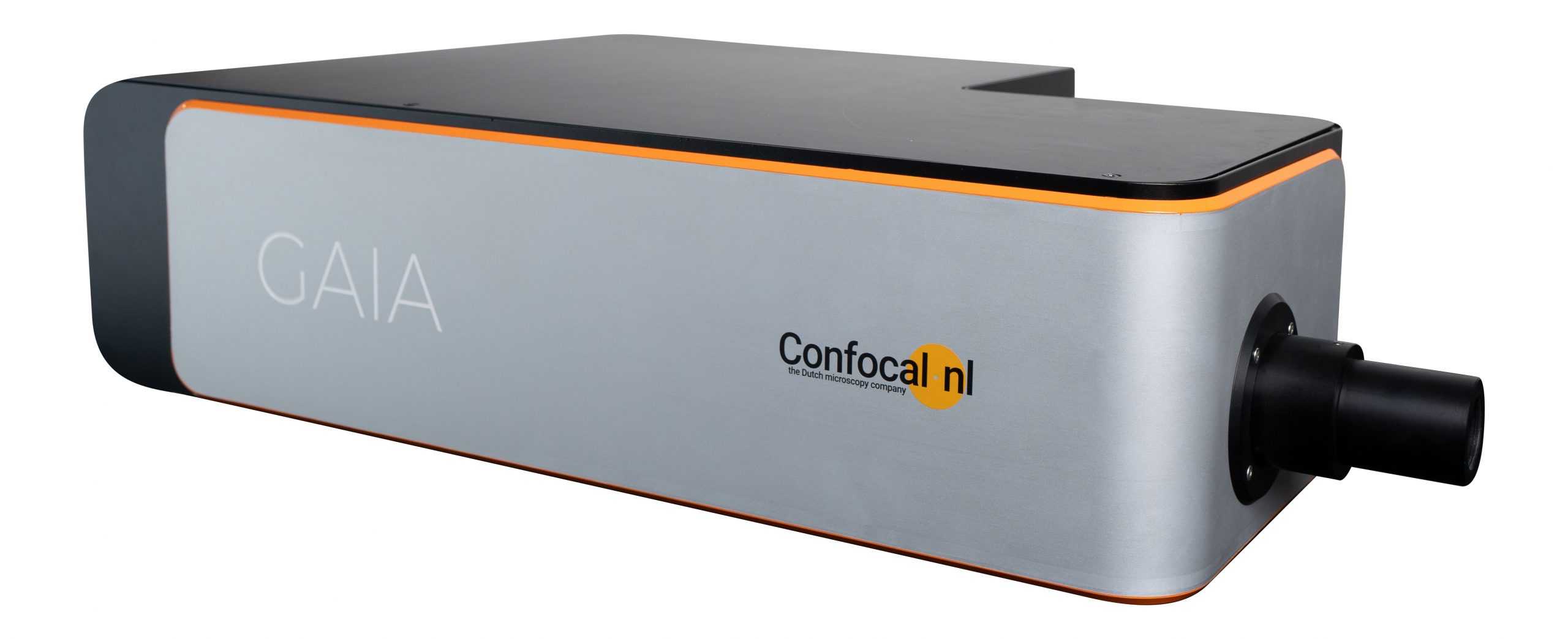
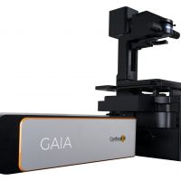
GAIA point-rescanning confocal microscope super-resolution, in a nutshell
Thanks to its patented rescanning confocal technology, GAIA point rescanning confocal is a super-resolution microscope enabling deep live cell imaging beyond the diffraction limit using only nanowatts of power. GAIA point-rescan is available in two versions, streamlined GAIA α and flagship GAIA λ.
GAIA point-rescanning confocal microscope, in more details
Imaging wide in super-resolution
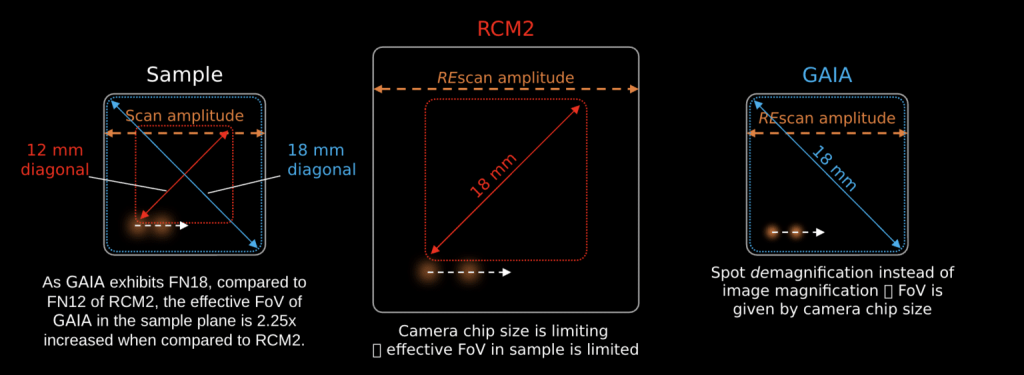
In laser rescanning confocal microscopy, the magnification of the image and the spot are decoupled. Thus, one can magnify the image compared to the spot. In RCM, we do this by giving the rescanners a larger amplitude than the scanners. As a consequence, all the details in the image are pulled further apart and the image is in super-resolution. However, this does not give infinite improvement in resolution. It turns out that the optimal situation is provided by twice as large rescan amplitudes, which improves the resolution by 1.4x.
In GAIA, instead of magnifying the image (through rescanning with higher amplitude), we demagnify the excitation and emission spot. Thanks to this revolutionary redesign of the rescan technology, GAIA provides more than double the field of view in the sample plane compared to its predecessor (RCM1 and RCM2). As a consequence, GAIA enables super-resolution imaging over a large field of view and using a wide range of objectives (30x -100x). For the best images high NA objectives are a must.
Imaging deep in super-resolution
Combination of low laser power requirements, high sensitivity of the detector and a novel optical design present in GAIA point rescan, enables super-resolution beyond 500 μm of depth. This makes GAIA point rescan confocal a perfect solution for super-resolution imaging of thick specimens like cleared tissue, whole zebrafish embryos, organoids and spheroids.
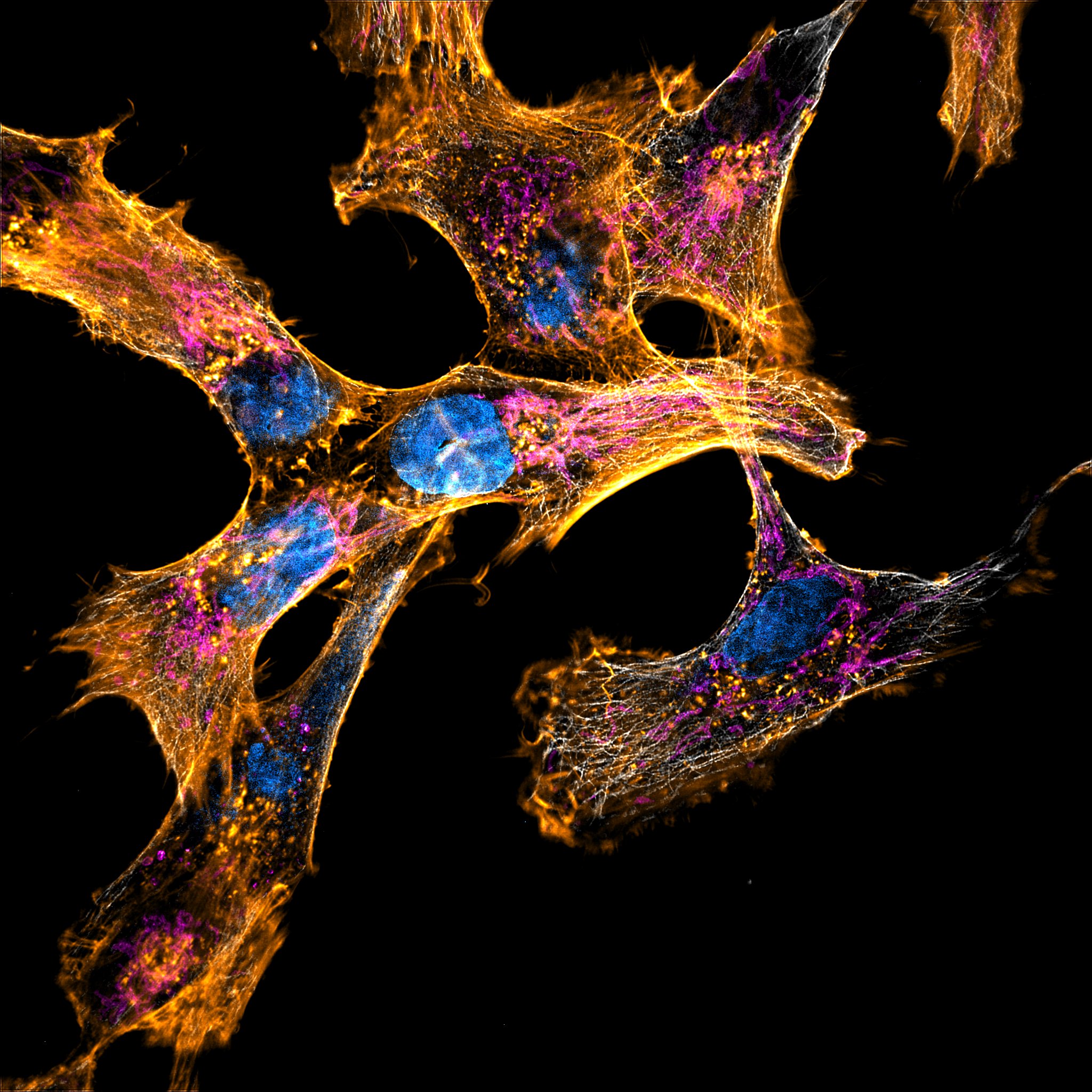
Imaging live in super-resolution
GAIA point rescan confocal is also ideal for live cell super-resolution imaging thanks to its very low photo-toxicity. The very low photo-toxicity of GAIA is attributed to two important factors:
- In traditional point-scanning confocal microscopy, high resolution is achieved by closing down the pinhole, reducing the number of photons collected by the detector. GAIA, like its predecessors RCM1 and RCM2, has the ability to achieve super-resolution while still using a large pinhole, thereby collecting more light.
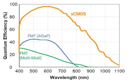
- The sCMOS and qCMOS cameras used in point rescanning confocal microscopy have much higher quantum efficiency than typical PMT detectors used in traditional point-scanning confocal microscopy, hence offering much higher sensitivity. This is illustrated by the curves below.
Imaging versatility in super-resolution
GAIA point rescanning confocal is the first system of its family to use a motorized switchable pinhole that enables perfect confocality for a wide range of objectives (4x up to 100x). For proper super-resolution, one should use high NA objectives.
To visit the manufacturer’s website, click HERE.
Applications of GAIA point-rescanning confocal microscopy:
Fast Cell Dynamics
- Better visualization and analysis of thin and dynamic structures even with dim signals
- Study fast cell dynamics with very low phototoxicity
- Cell migration
- Intra/extra-cellular transport
- Cell signaling
Live Cell Imaging
- Imaging happy cells and organisms for more than a day
- Long-term live cell imaging in gentle conditions
- Low phototoxicity and bleaching
- Developmental biology
- Regeneration
- Immunology
Deep 3D Imaging
- One slit pinhole for better images in deeper frames
- Organoids
- Zebrafish/mouse embryos
- 3D culture with hydrogels
White Papers:
Software compatible with GAIA point-rescanning confocal microscope:
Acquisition Software:
Both GAIAα and GAIAλ are compatible with Micro-Manager software for acquisition.
Deconvolution Software:
Both GAIAα and GAIAλ are compatible with Microvolution and SVI Huygens software for deconvolution.
Options for GAIA point-rescanning confocal super-resolution microscope:
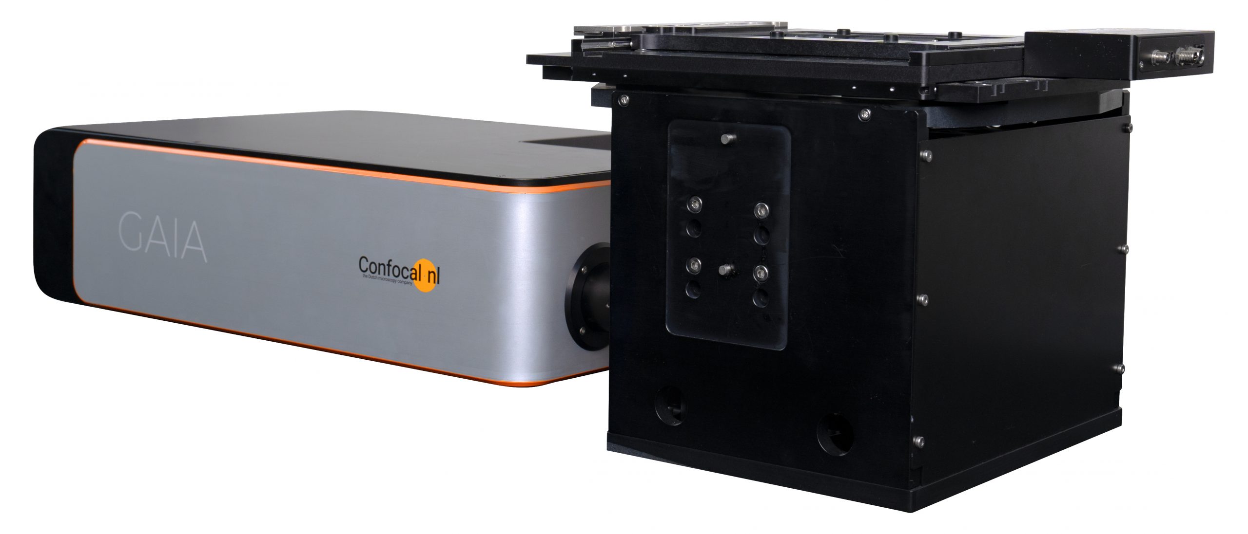
GAIAα and GAIAλ are confocal add-ons. To get a fully functional system a microscope stand, a camera and a laser launch are required. Below is a non-exhaustive list of devices compatible with GAIA systems.
Microscope Stands:
- Leica DMi8
- Nikon TE2000
- Nikon Ti-U & Ti-E
- Nikon Ti2-U & Ti2-E
- Olympus IX71 & IX81
- Olympus IX73 & IX83
- Zeiss Axio Observer.Z1
Cameras:
- Hamamatsu ORCA-Flash4.0 LT+, LT3 & V3
- Hamamatsu ORCA-Fusion & Fusion BT
- Hamamatsu ORCA-Lightning
- Hamamatsu ORCA-Fire & ORCA-Quest
Laser Combiners:
- Oxxius L4Cc & L6Cc