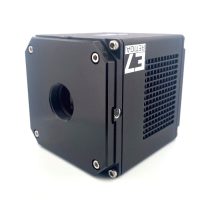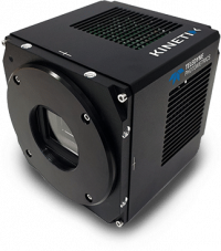


IRIS | Low Noise High Resolution sCMOS Camera
Sensor Technology
sCMOS
Quantum Efficiency
73%
Readout Noise
1.5 e-
Resolution
4.25 µm x 4.25 µm pixels
Speed
30 FPS
Interface
PCle, USB 3.0
 Description
 Specifications
 QE Curve
 Applications
 Downloads
Teledyne Photometrics Iris 9 & Iris 15 from Axiom Optics
High-resolution sCMOS cameras for wide-area imaging
The Teledyne Photometrics Iris family of high-resolution sCMOS cameras is designed for exceptional imaging at low magnifications, with fields of view up to 25 mm. Small 4.25 µm pixels spread across a large sensor capture fine detail over wide areas, giving a high-throughput solution without sacrificing image quality.
The Iris 9 offers 8.7 megapixels on a square sensor and the Iris 15 offers 14.9 megapixels across a 24.9 mm diagonal, both with 1.5 e- median read noise and 30 fps full-frame speed. Programmable Scan Mode synchronizes the rolling shutter to a scanned light sheet, making the Iris ideal for light-sheet microscopy, whole-slide imaging, high-content screening, and imaging large samples at high resolution. Axiom Optics supplies the Iris with application support for microscopy and imaging systems.
Interactive tool
Field of view, sampling, and tiling at low magnification
Small pixels are what let the Iris resolve fine detail through low-magnification objectives, while the large sensor covers more specimen per frame. Choose a model and objective to see the field of view, how many tiles a 10 x 10 mm sample needs, and whether the 4.25 µm pixels sample the optical resolution.
Near Nyquist
Close to ideal sampling at a low magnification, where larger-pixel cameras would discard detail. This is where the 4.25 µm pixel earns its place. An 11 µm pixel camera such as the Prime 95B would give 0.58 pixels per resolution element on this objective.
Near Nyquist
Close to ideal sampling at a low magnification, where larger-pixel cameras would discard detail. This is where the 4.25 µm pixel earns its place. An 11 µm pixel camera such as the Prime 95B would give 0.64 pixels per resolution element on this objective.
Nyquist sampled
About two pixels span each resolution element, so the Iris records everything this objective resolves across its full field. An 11 µm pixel camera such as the Prime 95B would give 0.77 pixels per resolution element on this objective.
Oversampled
The optics, not the pixels, now limit resolution. Use 2×2 binning to trade sampling for signal, or step down an objective to cover more of the sample per frame. An 11 µm pixel camera such as the Prime 95B would give 1.21 pixels per resolution element on this objective.
Oversampled
The optics, not the pixels, now limit resolution. Use 2×2 binning to trade sampling for signal, or step down an objective to cover more of the sample per frame. An 11 µm pixel camera such as the Prime 95B would give 1.24 pixels per resolution element on this objective.
Near Nyquist
Close to ideal sampling at a low magnification, where larger-pixel cameras would discard detail. This is where the 4.25 µm pixel earns its place. An 11 µm pixel camera such as the Prime 95B would give 0.58 pixels per resolution element on this objective.
Near Nyquist
Close to ideal sampling at a low magnification, where larger-pixel cameras would discard detail. This is where the 4.25 µm pixel earns its place. An 11 µm pixel camera such as the Prime 95B would give 0.64 pixels per resolution element on this objective.
Nyquist sampled
About two pixels span each resolution element, so the Iris records everything this objective resolves across its full field. An 11 µm pixel camera such as the Prime 95B would give 0.77 pixels per resolution element on this objective.
Oversampled
The optics, not the pixels, now limit resolution. Use 2×2 binning to trade sampling for signal, or step down an objective to cover more of the sample per frame. An 11 µm pixel camera such as the Prime 95B would give 1.21 pixels per resolution element on this objective.
Oversampled
The optics, not the pixels, now limit resolution. Use 2×2 binning to trade sampling for signal, or step down an objective to cover more of the sample per frame. An 11 µm pixel camera such as the Prime 95B would give 1.24 pixels per resolution element on this objective.
Resolution uses the Rayleigh criterion at 520 nm emission. Tile counts assume a 1x camera adapter, a field number large enough for the sensor, and no tile overlap; plan 10 to 15% more for stitching overlap.
Interactive visualizer
Programmable Scan Mode for light-sheet microscopy
A rolling-shutter sCMOS exposes a band of rows that sweeps across the sensor. Programmable Scan Mode opens up the sensor timing so that band can act as a virtual confocal slit, locked to a scanned light sheet: only rows inside the slit collect light, which rejects out-of-focus and scattered background. Choose a scan direction and slit width to see the timing.
The exposure window rolls from the top row to the bottom, following a light sheet that the scanner sweeps in the same direction.
The exposure window rolls from the top row to the bottom, following a light sheet that the scanner sweeps in the same direction.
The exposure window rolls from the top row to the bottom, following a light sheet that the scanner sweeps in the same direction.
Reversing the scan direction matches a sheet that sweeps upward, or a mirrored optical path, without remounting the camera.
Reversing the scan direction matches a sheet that sweeps upward, or a mirrored optical path, without remounting the camera.
Reversing the scan direction matches a sheet that sweeps upward, or a mirrored optical path, without remounting the camera.
Alternating frames read top to bottom, then bottom to top, so a bidirectional galvo can image on both strokes instead of waiting for flyback.
Alternating frames read top to bottom, then bottom to top, so a bidirectional galvo can image on both strokes instead of waiting for flyback.
Alternating frames read top to bottom, then bottom to top, so a bidirectional galvo can image on both strokes instead of waiting for flyback.
Animation slowed for display; slit drawn to scale. Exposure per row equals slit width times the line time, shown at the default 11.26 µs line time. Line Delay mode lengthens the line time to slow the sweep to the scanner, and Scan Width mode sets the slit directly. Programmable Scan Mode runs over PCIe.
Interactive explorer
Frame rate by model, interface, and region of interest
Both Iris models read one row every 11.26 µs, so over PCIe the frame rate depends only on how many rows are read. USB 3.0 is bandwidth limited, which matters most for the wider Iris 15 sensor.
Frame rates from the Teledyne Photometrics Iris 9 and Iris 15 datasheets, 16-bit.
Iris 9 vs. Iris 15
Both models use the GPixel GSense 5130 sensor with 4.25 µm pixels, over 73% peak QE, 1.5 e- median read noise, a 16-bit output, and a 200 to 1100 nm spectral range. They differ in sensor width, field of view, and lens mount.
| Model | Array | Sensor area | Diagonal | Full frame, PCIe / USB | Full well | Mount |
|---|---|---|---|---|---|---|
| Iris 9 | 2960 x 2960 (8.7 MP) | 12.61 x 12.61 mm | 17.8 mm | 30 / 16 fps | 10,000 e- | C-mount |
| Iris 15 | 5056 x 2960 (14.9 MP) | 21.49 x 12.61 mm | 24.9 mm | 30 / 10 fps | 13,000 e- | F-mount |
For high-magnification, photon-limited work, compare the Prime 95B with 11 µm pixels, or see the full scientific CMOS lineup.
Application guide
Where the Iris excels
Light-sheet and cleared-tissue microscopy
Light-sheet systems image whole organs, embryos, and cleared tissue volumes plane by plane. The Iris 15 covers a 25 mm field in one frame, its small pixels hold resolution at the low magnifications these systems use, and Programmable Scan Mode synchronizes the rolling shutter to the scanned sheet for confocal-like background rejection. Teledyne Photometrics customer stories include mesoSPIM volume imaging.
Whole-slide imaging and tiled acquisition
Every tile saved is time saved. A larger field of view per frame means fewer stage moves and stitching seams, and 4.25 µm pixels keep a 10x or 20x scan sharp enough for pathology and histology review.
High-content screening and cell counting
Screening throughput depends on how many cells fit in each field. The Iris captures more wells, colonies, or cells per exposure at low magnification while resolving the morphology that downstream analysis relies on.
Wide-field calcium and voltage imaging
Mapping activity across many neurons calls for a large field and enough resolution to separate neighboring cells. The Iris reaches 30 fps across its full sensor over PCIe and up to 695 fps on a 128-row region of interest.
Broadband and UV imaging
Teledyne Photometrics specifies the Iris spectral response from 200 to 1100 nm, so a single camera covers UV, visible, and near-infrared work. Axiom Optics can help pair it with UV optics and sources.
Key features
Resolution and pixel size
Small 4.25 µm pixels capture intricate detail across the imaging plane, giving excellent resolution with lower-magnification objectives.
sCMOS sensitivity
Low noise and high quantum efficiency across a broad wavelength range deliver better images with shorter exposures than traditional CCD cameras.
Advanced triggering
Programmable Scan Mode exposes sensor timing for precise line-time control, alongside up to four Expose Out signals and effective global shutter modes.
Superior background quality
Pattern Noise Reduction and Correlated Noise Reduction technologies produce clean, pattern-free images with minimal pixel defects, even in low light.
Compact form factor
At 76 x 76 x 88 mm for the Iris 9 and 78 x 78 x 108 mm for the Iris 15, with optimized air cooling, the Iris fits new and existing setups.
PCIe or USB 3.0
Choose PCIe for full speed and Programmable Scan Mode, or USB 3.0 for simple plug-in integration. Supported by the PVCAM SDK, Ocular, and third-party software.
Iris frequently asked questions
What is the Iris sCMOS camera?
The Iris from Teledyne Photometrics is a family of high-resolution scientific CMOS cameras built for wide-area imaging. The Iris 9 has a 2960 x 2960 array and the Iris 15 a 5056 x 2960 array, both with 4.25 µm pixels on the GPixel GSense 5130 sensor and fields of view up to 25 mm. Axiom Optics supplies and supports both models.
What is the difference between the Iris 9 and Iris 15?
Both share the same pixels, QE, read noise, and 30 fps full-frame speed over PCIe. The Iris 15 has a wider 21.49 x 12.61 mm sensor with a 24.9 mm diagonal and an F-mount, while the Iris 9 has a square 12.61 x 12.61 mm sensor with a 17.8 mm diagonal and a C-mount. The Iris 15 also has a deeper 13,000 e- full well versus 10,000 e- for the Iris 9.
Why choose small 4.25 µm pixels?
Pixel size at the sample equals camera pixel size divided by magnification. Small pixels reach Nyquist sampling at lower magnifications, about 20x with a 0.75 NA objective, so you can use low-power objectives that see more of the sample per frame without losing resolution.
Is the Iris suitable for light-sheet microscopy?
Yes. Programmable Scan Mode lets the rolling shutter act as a virtual slit synchronized to a scanned light sheet, with selectable scan direction, slit width, and line delay. Combined with a field of view up to 25 mm, this makes the Iris a strong fit for light-sheet, cleared-tissue, and mesoscale imaging. Programmable Scan Mode requires the PCIe interface.
How fast are the Iris cameras?
Both models run at 30 fps full frame over PCIe, rising to 59 fps at 1500 rows, 174 fps at 512 rows, and 695 fps at 128 rows. Over USB 3.0 the Iris 9 reaches 16 fps and the Iris 15 reaches 10 fps at full frame.
What are the Iris sensitivity specifications?
Both models offer over 73% peak quantum efficiency, 1.5 e- median read noise, and a 16-bit output, with spectral response from 200 to 1100 nm. They are air cooled to 0 °C at 30 °C ambient with 0.5 e-/pixel/second dark current.
How many images does it take to tile a large sample?
With a 4x objective, the Iris 15 covers 5.37 x 3.15 mm per frame and needs 8 tiles for a 10 x 10 mm sample, while the Iris 9 needs 16 tiles, before allowing for stitching overlap. Fewer tiles mean faster scans and fewer seams.
When should I choose the Prime 95B instead?
Choose the Prime 95B for photon-limited imaging at high magnification, such as single-molecule work or spinning disk confocal at 60x to 100x, where its 11 µm pixels and 95% QE collect more signal per pixel. Choose the Iris for resolution and field of view at low magnification.
Choose the right Iris for your system
Axiom Optics imaging specialists can match the Iris model, interface, and lens mount to your microscope or light-sheet setup and help configure Programmable Scan Mode timing.