
 Description
 Specifications
 Applications
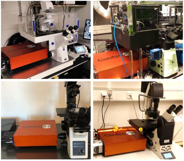
 Pictures
 Videos
 Downloads
 Publications
What is RCM?
RCM stands for rescan confocal microscopy. It’s also a sensitive, high resolution and affordable confocal imaging system:
- An ideal solution for small labs with limited budget, but demanding tasks, particularly when high sensitivity and resolution are desired from the imaging system,
- A scanning confocal microscope that works as a camera, no need for an instruction manual
- RCM is extremely easy to use: no hardware control or software processing needed, and the images are always RAW.
RCM can be delivered as a total microscope system with a selection of microscopes ( Nikon, Olympus, Leica or Zeiss ), a selection of cameras ( Hamamatsu, PCO, Andor, Photometrics ) and laser solutions (Omicron, Toptica ).
In case you already have a microscope in the lab, RCM is an upgrade to an existing wide-field fluorescence system – RCM can easily be added to the existing wide-field fluorescence microscope system to improve its resolution.
RCM Working Principle
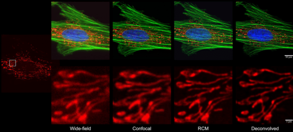
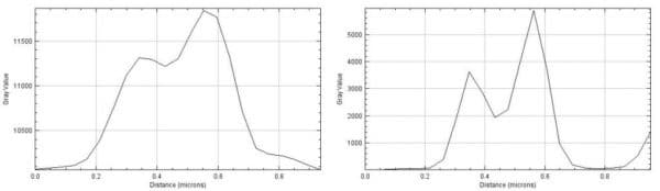
The RCM technique extends standard confocal microscopy with a re-scanning unit, improving lateral resolution by √2 and reducing signal to noise ratio.
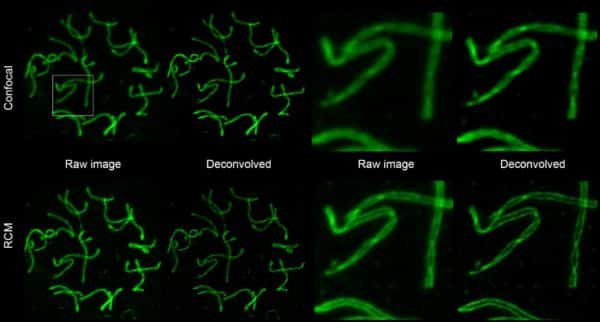
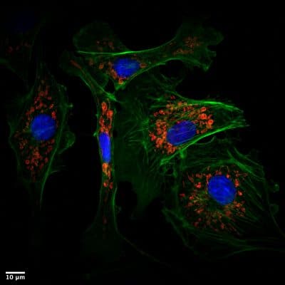
Re-scan Confocal Microscopy (RCM) is a new super-resolution technique based on standard confocal microscopy extended with an optical (re-scanning) unit that projects the image directly on a CCD-camera. This new microscope has improved lateral resolution (170 nm at 488 nm excitation), and strongly improved sensitivity, while maintaining the sectioning capability of a standard confocal microscope. It is particularly useful for biological applications where the combination of high-resolution and high-sensitivity is required (but not very high imaging speed).
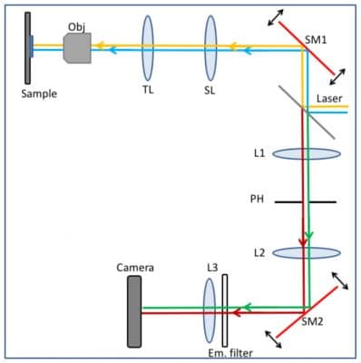
The excitation lasers (blue and yellow lines) are directed via a dichroic mirror towards the first scanning unit SM1. As in a standard confocal microscope, the scanning unit scans the laser light in the sample and de-scans the emission light, directing it at the pinhole PH (green and red lines). After the pinhole, a second re-scan unit SM2 directs the light onto a camera chip.[/caption]
During scanning, re-scan mirrors (SM2) move faster than the first scan mirrors (SM1). This magnifies the image on the camera chip compared to the sample, and eventually results in the higher resolution of the image. The resolution of the system is improved with the re-scan step by a factor of √2 (i.e. 1.41 times), compared to Abbe’s resolution limit by changing the angular amplitude of the re-scanner (SM2). Reduction of pinhole is no longer necessary to increase resolution. Closing down the pinhole only limits the amount of light passing through and decreases the signal to noise ratio due weaker signal. Since the re-scan is a purely optical method with no further image processing required, there is cost in time while improving the resolution. By using a sensitive camera as detector, the signal-to-noise ratio of the RCM is 4 times higher than in standard confocal microscopy.
To fully understand the principle of rescanning, resolution improvement and the optical layout of the RCM, please watch the video below that explains the components and the light path of the RCM (animation credits to StudioFlip). Additional technical details and test images can be found in De Luca et al (2013).
Watch the working principle here
The Re-scan Confocal Microscopy (RCM) module can be used to turn any fluorescent microscope into a confocal microscope. For an upgrade, laser(s) and a camera are needed.
Check magnificent Turnkey examples from our customers for your inspiration:
- Andreas Kurz from Markus Sauer lab (University of Würzburg)
- Aymeric d’Hérouël from the Luxembourg Centre for Systems Biomedicine
Let us know which equipment you have available, and we will make a custom tailored upgrade solution for you.


 Working Principle