


 Description
 Specifications
 Applications
 Downloads
 Publications
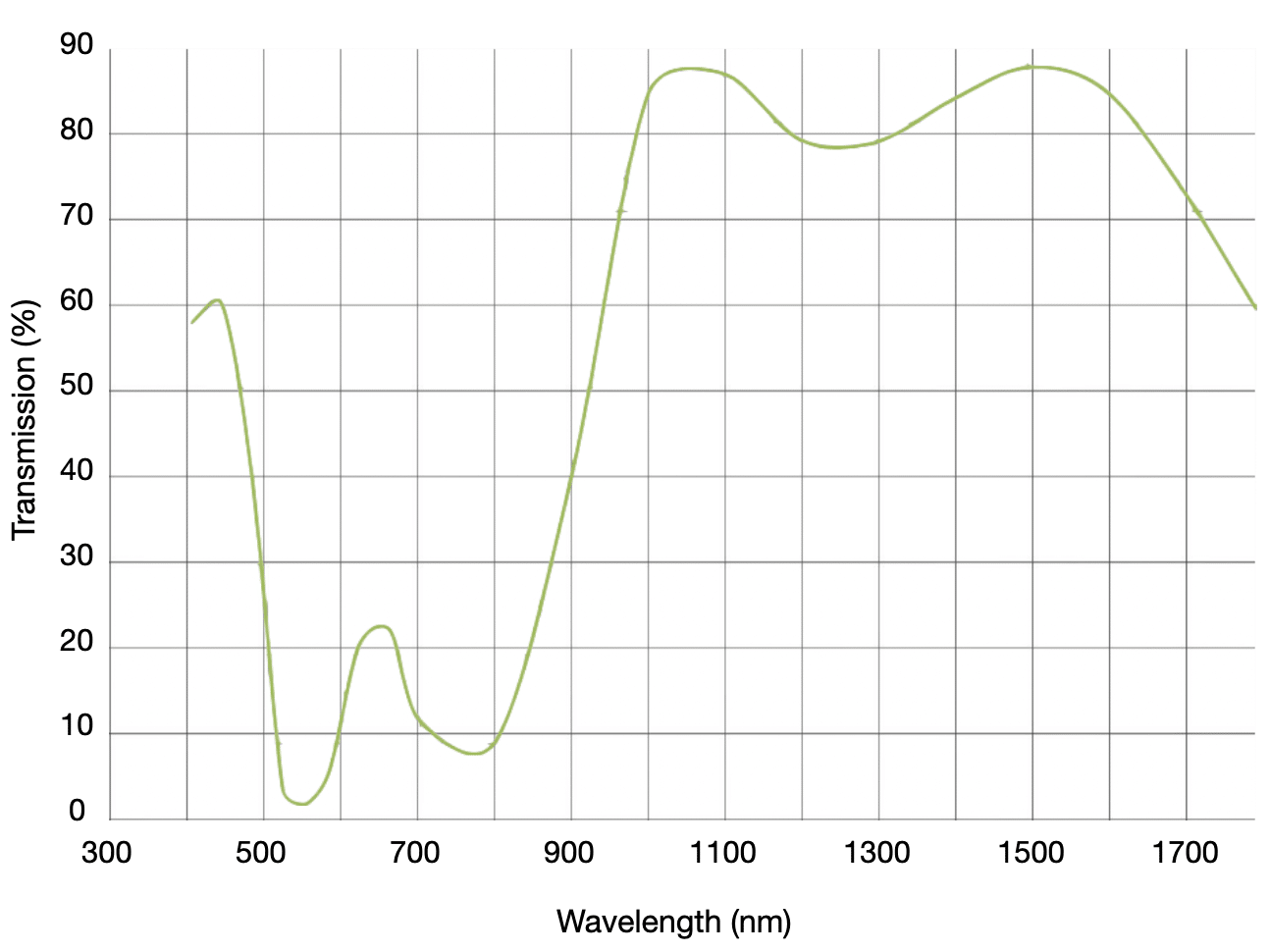

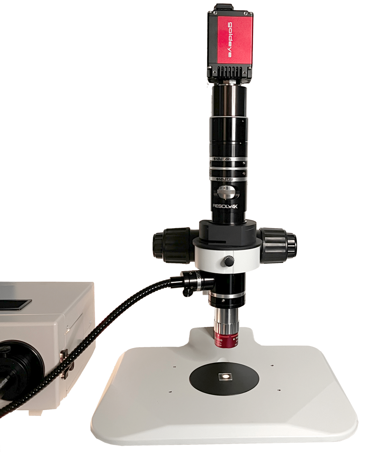
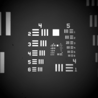
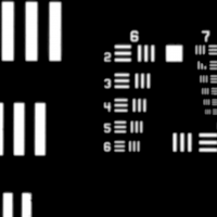
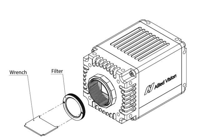
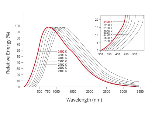
The HD-SWIR Digital Microscope is designed to “see” beyond what a standard optical microscope can image. Combining SWIR coated optics, SWIR illumination and a high resolution SWIR camera, the HD-SWIR microscope makes SWIR microscopy a cost effective solution that can image microscopic samples in the visible and the shortwave infrared region, thus covering a spectral range from 400 to 1700 nm. A variety of filters allows imaging in the SWIR only 0r to focus on spectral band or wavelength of interest.
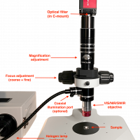
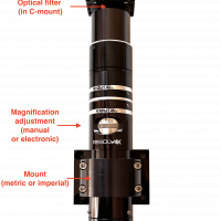
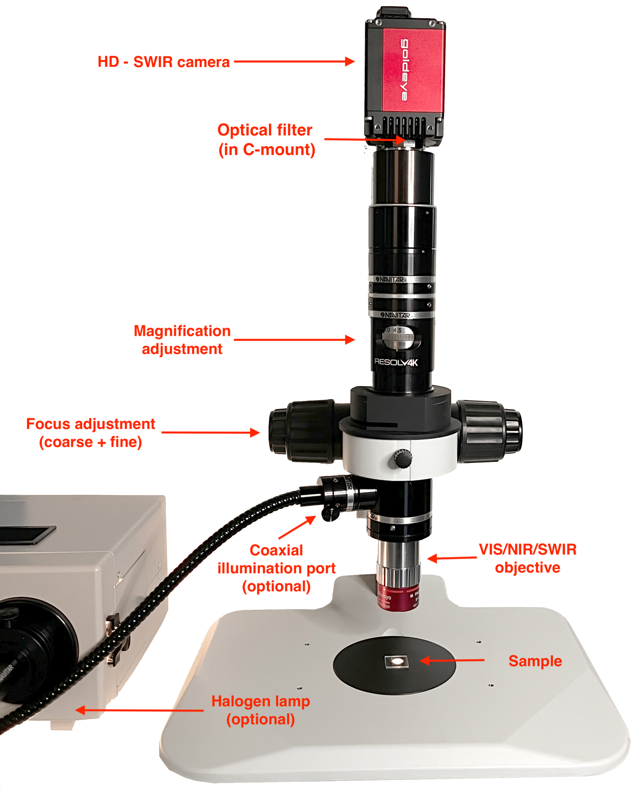
HD-SWIR Digital Microscope main components
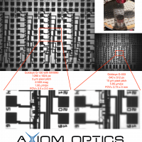
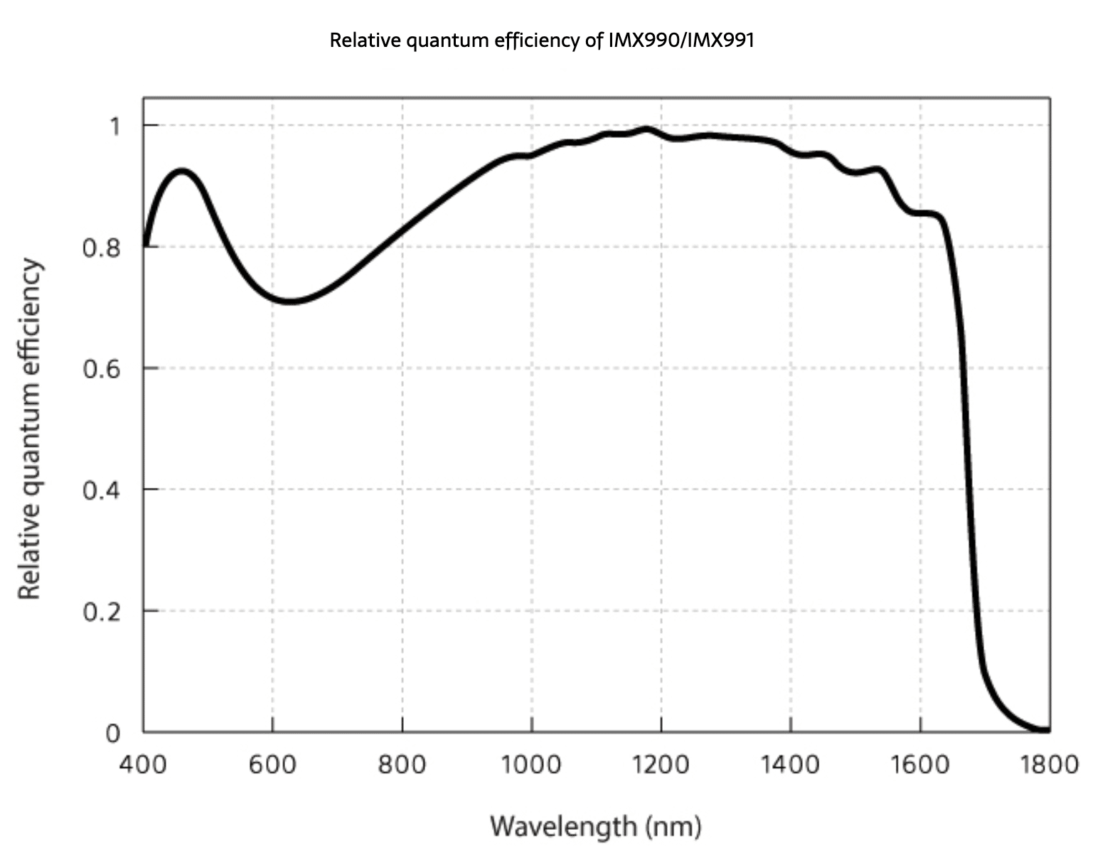
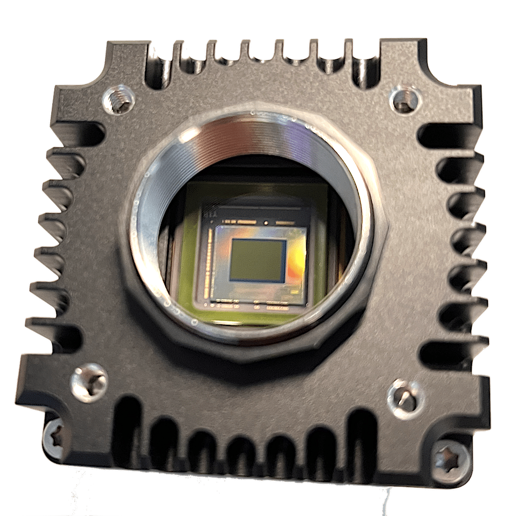
- A high-resolution SWIR InGaAs camera with 1280 x 1024 pixels (or 656 x 512), 5 µm pixel pitch, and high QE from 400 to 1700 nm. The 5 micron pixel pitch is the smallest in the industry, thus enables resolutions never reached before for SWIR wavelengths. A visualization software and complete SDK allow for easy use in the lab (tabletop version) or in production environment (industrial version). Other camera models (deep cooled or high-speed) are available upon request (see SWIR camera page here). The Goldeye G-130 uses the IMX990 and enables high resolution SWIR microscopy
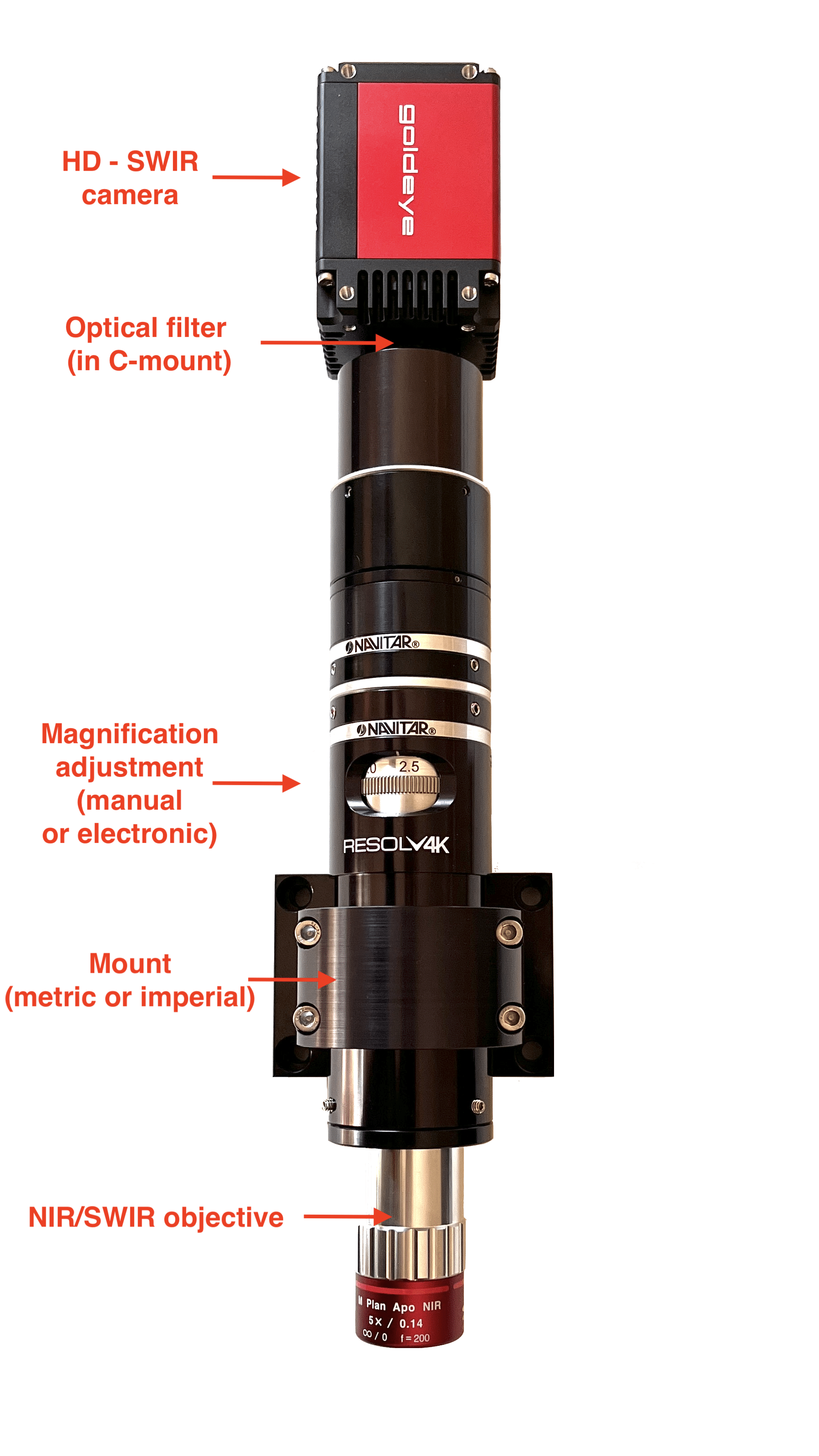
- SWIR optimized optics: all optics, included the microscope objective, are coated for optimal transmission in the SWIR (>900 nm). Magnification can easily be adjusted manually or electronically, thus allowing to quickly adapt the field of view.
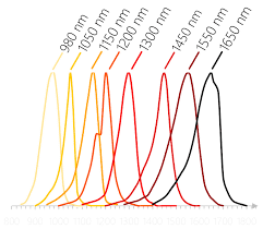
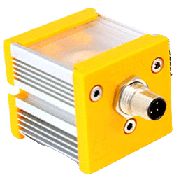
- SWIR illumination: a halogen lamp (broad spectrum covering visible and SWIR) can be used with a coaxial port, thus allowing illumination of the sample directly through the optics. Optionally, transmission-mode imaging is possible using a SWIR LED backlight with single wavelength emission spectrum.
Tabletop and industrial designs
-
- the tabletop design includes a stand with focus adjustment (coarse + fine) and an optional X/Y translation mount for holding the sample
- the industrial design allows easy integration thanks to a clamp mount. The long working distance of the objective (>30 mm) facilitates sample manipulation while maintaining a safe distance.
For more SWIR applications, visit our application page.
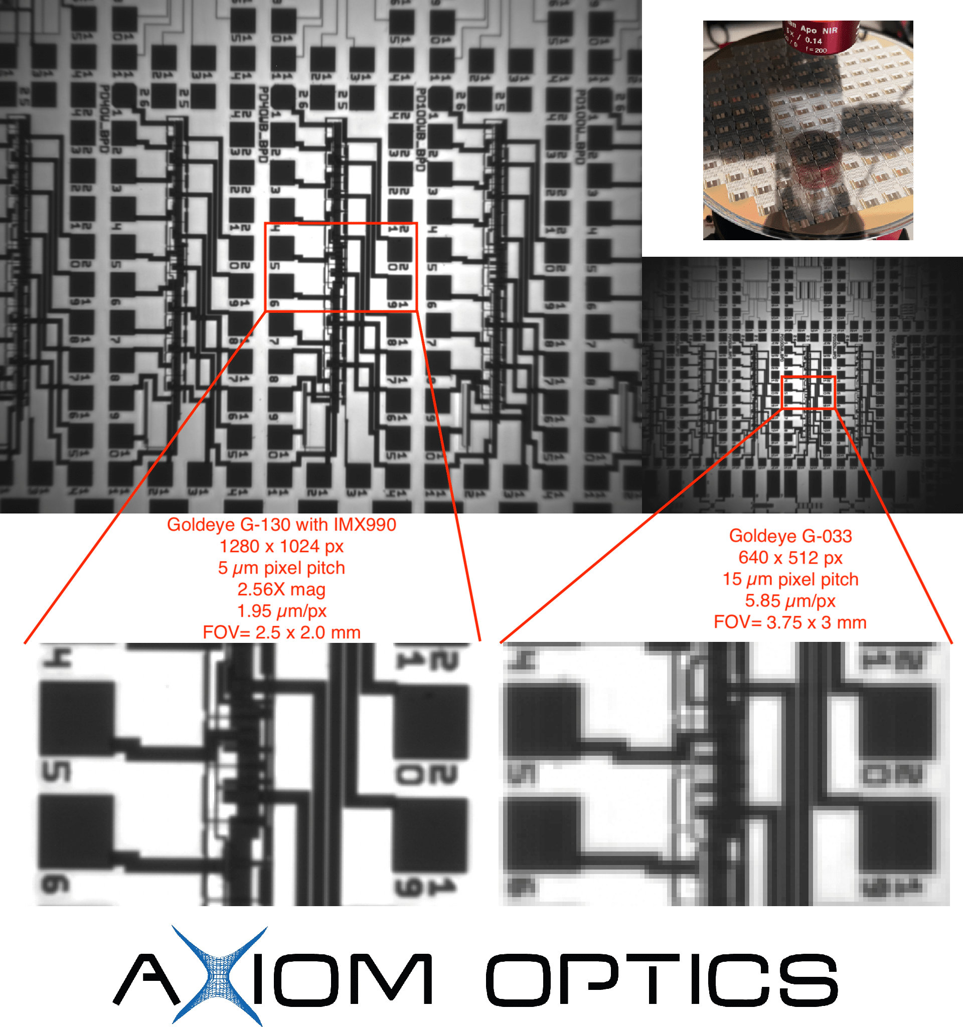
wafer inspection using HD-SWIR microscope and IMX990 ingaas camera