Introduction
Microscopes are essential to nearly every field of material and life science. The ability to observe the cells of living things, the structures of crystals, and the chemical composition of substances have been an enormous leap forward in scientific exploration.
Microscopes have evolved from crude assemblages of lenses and eyepieces to sophisticated instruments that can discern different structures at the molecular level. By examining how a substance or organism reacts to light through absorption or reflection (optical microscopy), investigators can identify diseases, examine how medicines affect cells, and more.
Widefield fluorescence and confocal fluorescence microscopy are two commonly used techniques that will reveal samples differently and will answer different scientific questions.
Composition of a Compound Fluorescence Microscope
Modern compound microscopes are complex pieces of machinery, and their main goal is to magnify an object of interest and create a 2D image that you can observe. The sample is illuminated by a light source, and the observer can look at the back-reflected or the transmitted light. Those are widely used techniques, but the contrast is not optimal as light comes from all parts of the sample, and therefore, it’s hard to distinguish fine structures.
Fluorescence microscopy is a technique that drastically improves contrast and is extremely popular in cell biology and molecular biology. Fluorescence is a phenomenon by which a chemical compound (fluorophore) can re-emit light upon light excitation. The emitted light is of a longer wavelength than the excitation light, so by using a proper set of optical filters, we can distinguish the emitted signal from the excitation signal. By binding those fluorophores to specific structures of a tissue or cell, we can observe this structure without collecting light from the other parts of the tissue or cell. Contrast is highly improved as only the labeled structure will emit light.
The main parts of a fluorescence microscope include the following:
Excitation Light Source
Historically, tungsten-halogen, mercury, or xenon arc-lamp have been largely replaced by LEDs and lasers. A single LED emits a spectrum several tens of nanometers wide, so combining several LEDs can cover the whole visible spectrum. Lasers have much narrower excitation bands, typically one nanometer or less.
Filter Turret
As an assembly of an excitation filter, a dichroic mirror and an emission filter select the specific excitation and emission lights of a fluorophore. Each part plays an important role, as described below.
Excitation Filter
The excitation filter blocks all but the specific wavelengths required to excite a specific fluorophore. For instance, a GFP excitation filter will let light centered around 470 nm go through and will cut the rest of the spectrum. Lasers do not require excitation filters as they are already highly specific.
Dichroic Mirror
The excitation light then bounces off a dichroic mirror toward the sample. The dichroic mirror is designed to reflect some parts of the spectrum and let some other parts go through. For instance, a GFP dichroic mirror will reflect light below 490 nm, and light above 500 nm will be transmitted.
Emission Filter
The emission filter blocks all but the specific wavelengths emitted from a specific fluorophore. For instance, a GFP emission filter will let light centered around 525 nm go through and will cut the rest of the spectrum.
Objective
The objective focuses the excitation light reflected from the dichroic mirror to the sample, collects the emission signal emitted from the sample, and sends it back to the dichroic mirror. The objective is an assembly of lenses housed within a circular casing that will magnify the image up to 100 times.
Stage
The stage is the platform where the sample or specimen rests for observation under the microscope. The stage can move in the lateral or axial directions and can be motorized for more complex acquisitions (z-stacking, xy-tiling/stitching, etc.).
Detector
The director is where the emission light from the sample terminates its travel. Photons are captured and generate electrons, which are then recorded to create a digital image of the sample.
How Do Widefield Microscopes Work?
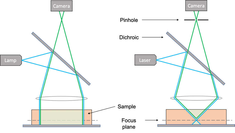
Widefield microscopes use LEDs for the excitation light source, and the entire volume below the objective lens is illuminated in a homogeneous way. This means every fluorophore contained in that volume is likely to emit photons and contribute to the emission signal. This technique is not axially selective, as all axial planes are contributing, which creates a brighter but blurrier image.
How Do Confocal Microscopes Compare?
Confocal microscopes use lasers for the excitation light source. As lasers are already very selective light sources, no excitation filter is required, but the dichroic mirror and emission filter can still be required. In a confocal microscope, the light is focused down to the focal plane of the objective lens. Because the energy of the laser is focused on a small volume of the sample, mostly the fluorophores contained in that small volume will emit photons. This is highlighted in the schematics below:
This technique is very selective in the lateral direction. The focused beam scans through the focal plane to create a 2D image. Every fluorophore contained in that focused volume will sequentially emit photons, which will lead to the reconstruction of a 2D image. The fluorophores below and above the focal plane have contributed much less than the fluorophores in the focal plane, but their contribution is not zero. Confocal microscopes use another component to improve even more the axial selectivity: a pinhole. A pinhole is a tiny hole positioned in a conjugated plane to the objective lens that blocks all the out-of-focus light, only letting light from the focal plane go through. This is highlighted in the schematics below:
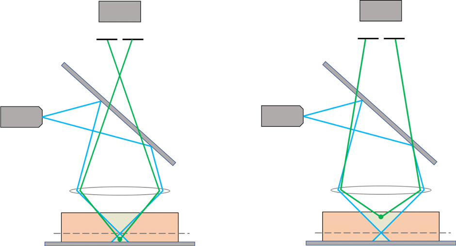
The result is a sharp and highly focused image. By axially translating the stage and recording more images, we can then reconstruct a 3D volume of the sample.
Advantages and Drawbacks of Each Technique?
Axial Sectioning
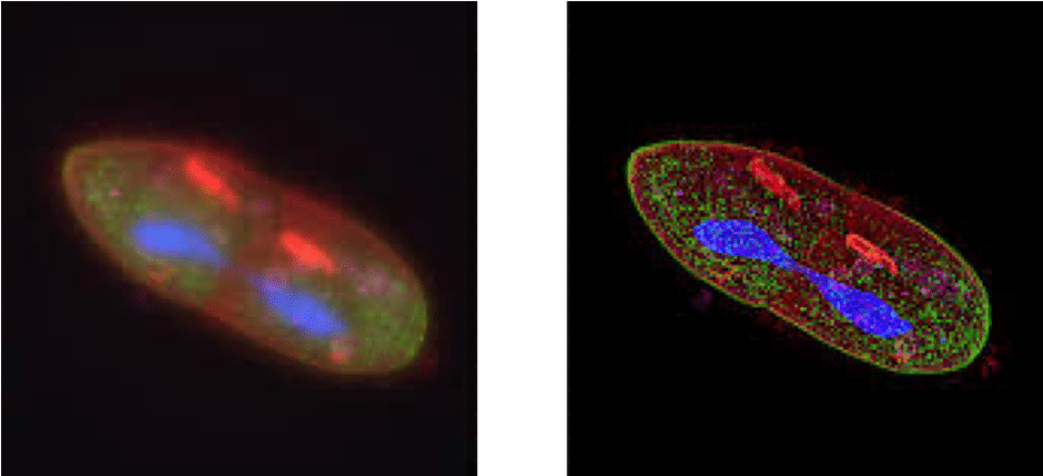
As explained before, the main advantage of confocal microscopy is to get axially selective images and reconstruct them in 3D with fewer axial artifacts than in standard widefield microscopy. An image of a sample acquired with a confocal microscope will always look sharper than the same sample acquired with a widefield microscope (see example below).
Resolution
Confocal microscopes, thanks to the use of a pinhole, slightly improve the lateral resolution compared to widefield microscopes. Furthermore, there are a number of techniques to improve the resolution even further, like the rescan technique, which is used to achieve super-resolution with the RCM1 or RCM2. You can find more information here.
Sensitivity/Phototoxicity
But because a widefield microscope captures photons emitted from a much larger volume, the signal intensity is usually much brighter than on a confocal microscope. Therefore, they don’t need as intense light sources and are less phototoxic.
Speed
Widefield microscopes are also faster than traditional laser-scanning confocal microscopes. When combined with their higher sensitivity, they are great choices for live cell imaging to capture fast-moving biological events and/or organisms. Spinning-disk and line-scanning confocal microscopes (like the NL5) are high-speed confocal systems that can be used for applications requiring both confocal imaging and high temporal resolution.
Price
Confocal microscopes are usually significantly more expensive as they require high-precision optics and mechanics and fancier light sources.
Overall, which technique you use depends on the nature of the sample and of the inquiry. If you are imaging a very flat sample (less than a couple of µm thick), chances are that a widefield microscope will be sufficient, but if you need better resolution, sectioning, and contrast, then a confocal microscope will prove to be a very useful system.