Forget the monochrome world of black-and-white, and even the simple RGB canvas of a traditional color image. Hyperspectral Microscopy is the technology that moves imaging into the 21st century by treating every single pixel not just as a spot of color, but as a treasure trove of spectral data.
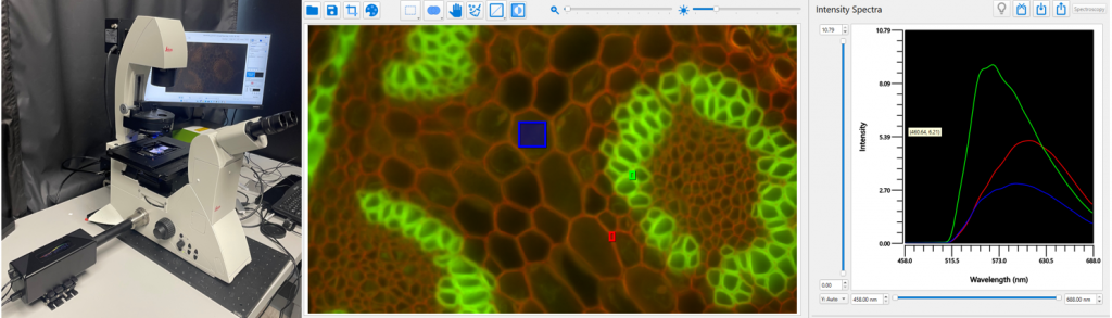
Look into the eyepieces of your microscope and see hundreds of distinct colors at once. That’s the power of Hyperspectral Microscopy: it combines high-resolution microscopy with spectral analysis, capturing data across tens to hundreds of spectral bands at every pixel. A standard image becomes a multidimensional data cube.
The Invisible Revealed
In traditional imaging, two different materials that share the same color might look identical. But with Hyperspectral Microscopy, one can analyze the unique spectral signature (a material’s “optical fingerprint”) pixel-by-pixel. It is possible to differentiate:
- Tissue Types: Subtle differences in cellular structure or chemical composition are instantly apparent.
- Chemical Compounds: Distinguishing between different polymers or drug states.
- Cell States: Identifying changes in cell health or function.
This label-free capability allows for the integrity of sensitive biological samples to remain intact for long-term or in vivo studies. For example, in a porcine model, Hyperspectral Imaging has been used during pancreatectomy to precisely and non-invasively quantify pancreas perfusion in real-time.
Beyond Simple Pictures
Hyperspectral Microscopy doesn’t just offer better pictures; it offers better data. This richness translates directly into enhanced sensitivity and specificity.
Hyperspectral imaging detects minute spectral differences—like the shift in a single protein’s conformation—that could be missed by conventional modalities. This is critical for early disease detection, such as pinpointing precancerous tissue changes before they become obvious. In materials science, it offers non-destructive, detailed characterization of everything from nanoparticles to complex composite polymers.
Because the data is so rich, Hyperspectral Microscopy is tailor-made for the age of AI and machine learning. It turns what used to be subjective, qualitative interpretation into data-driven insights that are repeatable and easily scalable for automated classification.
A prime clinical application is the extraction of oxygen blood content from the spectral data cube. Unlike current fluorescence angiography, which requires injecting exogenous fluorophores and provides non-quantitative results, Hyperspectral Microscopy offers a relatively easy, non-invasive, and quantitative path to monitoring tissue oxygenation.
The Trade-Off
There are no free lunches, there is always a price to be paid somewhere and for Hyperspectral Microscopy, it’s speed. Since the system divides the available light (photons) into many narrow spectral bands, each individual band receives less light. This means hyperspectral images can take longer to acquire, or require more intense light, to achieve an acceptable signal-to-noise ratio. There are other trade-offs as well to get the hyperspectral data: manufacturers sacrifice resolution, field of view as well as acquisition speed to gather all this data.
In summary, Hyperspectral Microscopy offers sensitivity, specificity, and multiplexing, all while enabling label-free, quantitative, and non-destructive imaging. These advantages are driving rapid adoption, positioning it as a truly transformative technology in modern science and medicine.
Ready for Your Upgrade?
Axiom Optics offers several ways to integrate a spectral solution into your lab:
- Silios (Snap-Shot Multiplexing): Like a specialized digital camera, Silios uses a Bayer-like filter pattern on the sensor to capture up to 10 optical bands simultaneously. This technique is fast, with cameras covering the visible spectrum through SWIR.
- Hinalea (The Scanner): Hinalea uses a tunable Fabry-Perot cavity to sequentially scan through hundreds of spectral bands for each image. This provides the highest spectral resolution, with sensors ranging from UV to XSWIR.
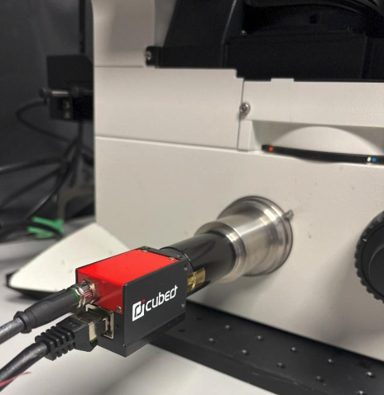
- Cubert (Light-Field Approach): Cubert divides the sensor into sections, where each section captures data from a single optical band. There is no loss of spatial resolution—only field of view. They offer up to 64 optical bands with sensors sensitive in the VIS, NIR, and SWIR.
To learn more about our Hyperspectral Camera Portfolio click HERE
This post was written by:
Scott Phillips, Sr. Technical Sales Engineer