Light Microscopy
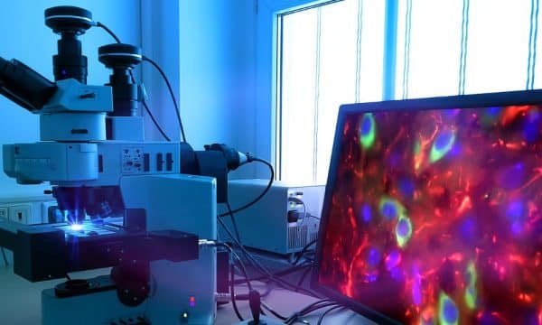
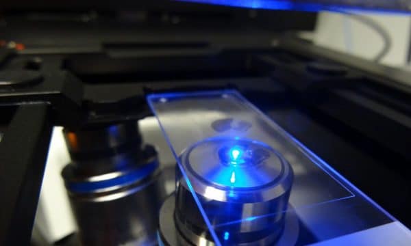
Light microscopy is a critical tool in modern cell biology and neurosciences. Contrary to an electron microscope, a light microscope uses light to image small objects. The relatively gentle nature of light means that living organisms or cells can be observed for long periods of time and their dynamics can be studied extensively.
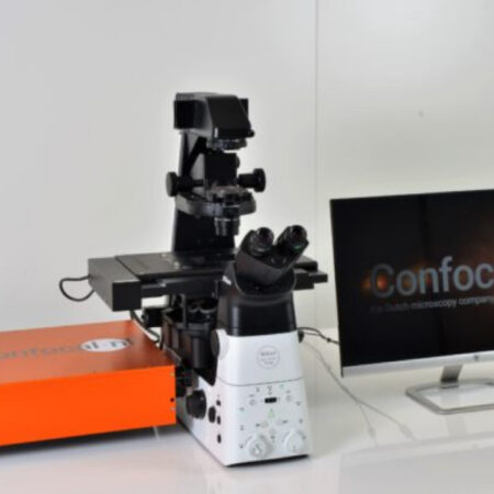
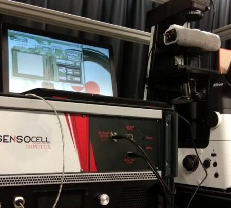
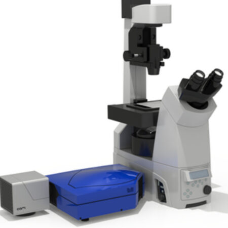
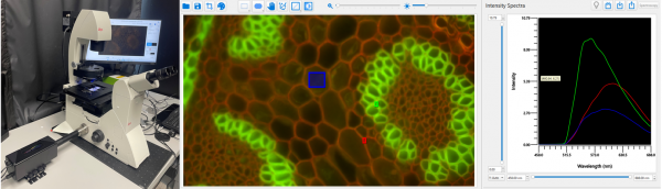
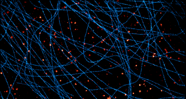
Axiom Optics offers a variety of high end light microscopy instruments, including add-ons for confocal imaging, super-resolution imaging, fluorescence lifetime Imaging (FLIM), TIRF imaging, optical tweezers for mechanobiology, adaptive optics, single-molecule localization and Argolight slides for calibration and microscope metrology.
Contact Axiom Optics to discuss your light microscopy needs. One of our experts will help you select the right system, satisfying all your imaging requirements.
Read More