
Green lasers, specifically 532 nm CW lasers, are foundational tools across a diverse range of scientific, industrial and medical applications. Their prominence stems from a unique combination of highly desirable characteristics: high beam quality, a visible green output, stable output power, and strong absorption in many materials. Together, these attributes make 532 nm CW lasers indispensable for both delicate diagnostic procedures and robust, demanding industrial processes.
One of their most valuable features is the ability to efficiently generate both fluorescence and Raman signals, which makes them central to spectroscopy, bioimaging and materials analysis. In spectroscopy, the strong interaction of 532 nm light with many molecules allows precise identification and quantification of chemical components. In bioimaging, efficient excitation of fluorescent markers enables high-resolution visualization of cellular and subcellular structures. In materials analysis, the distinctive Raman scattering generated by 532 nm light offers a non-destructive way to determine the chemical composition and structure of materials ranging from semiconductors to pharmaceuticals.
High beam quality
TEM00 output with low noise and stable power for precise measurement.
Visible green
Easy alignment and high visibility for displays and inspection.
Strong interaction
Efficient Raman and fluorescence excitation, and strong absorption by hemoglobin and melanin.
Raman Spectroscopy: A Cornerstone Application for 532 nm Lasers
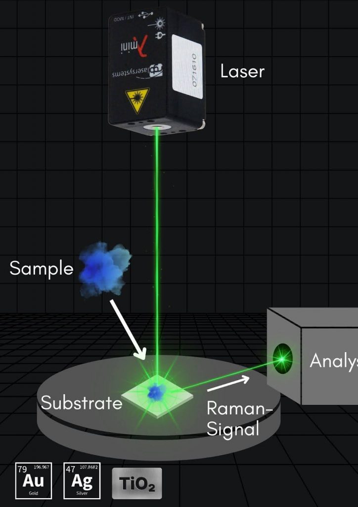
One of the most significant uses of 532 nm CW lasers is Raman spectroscopy, a non-destructive analytical technique that identifies molecular structures from their vibrational energy modes.
- Strong Raman signal excitation: 532 nm strikes a balance between signal intensity and fluorescence interference, ideal for inorganic materials and many organic ones.
- Versatility across sample types: it is widely used to study nanomaterials such as graphene and carbon nanotubes, semiconductors, biological samples and pharmaceutical compounds.
- Portable and benchtop systems: compact CW green lasers enable handheld Raman instruments for security screening, art conservation and quality control.
Where does the Raman signal land?
Pick an excitation wavelength and a Raman band to see the scattered wavelength, relative signal and detector fit.
The Silicon band lands at 547.1 nm, well inside the silicon detector range.
Highest Raman efficiency, but the strongest risk of fluorescence background in colored or biological samples.
The Graphene D band lands at 573.2 nm, well inside the silicon detector range.
Highest Raman efficiency, but the strongest risk of fluorescence background in colored or biological samples.
The Graphene G band lands at 580.8 nm, well inside the silicon detector range.
Highest Raman efficiency, but the strongest risk of fluorescence background in colored or biological samples.
The Graphene 2D band lands at 621.2 nm, well inside the silicon detector range.
Highest Raman efficiency, but the strongest risk of fluorescence background in colored or biological samples.
The C-H stretch band lands at 629.0 nm, well inside the silicon detector range.
Highest Raman efficiency, but the strongest risk of fluorescence background in colored or biological samples.
The O-H stretch band lands at 649.5 nm, well inside the silicon detector range.
Highest Raman efficiency, but the strongest risk of fluorescence background in colored or biological samples.
The Silicon band lands at 818.4 nm, well inside the silicon detector range.
A common compromise: much less fluorescence than 532 nm, with lower Raman efficiency.
The Graphene D band lands at 878.1 nm, well inside the silicon detector range.
A common compromise: much less fluorescence than 532 nm, with lower Raman efficiency.
The Graphene G band lands at 896.1 nm, well inside the silicon detector range.
A common compromise: much less fluorescence than 532 nm, with lower Raman efficiency.
The Graphene 2D band lands at 996.1 nm, well inside the silicon detector range.
A common compromise: much less fluorescence than 532 nm, with lower Raman efficiency.
The C-H stretch band lands at 1016.4 nm, near the long-wavelength edge of silicon detectors, where sensitivity drops.
A common compromise: much less fluorescence than 532 nm, with lower Raman efficiency.
The O-H stretch band lands at 1070.8 nm, near the long-wavelength edge of silicon detectors, where sensitivity drops.
A common compromise: much less fluorescence than 532 nm, with lower Raman efficiency.
The Silicon band lands at 1126.3 nm, beyond the silicon detector range; an InGaAs detector is needed.
Lowest fluorescence, but the weakest Raman signal and an InGaAs detector.
The Graphene D band lands at 1242.5 nm, beyond the silicon detector range; an InGaAs detector is needed.
Lowest fluorescence, but the weakest Raman signal and an InGaAs detector.
The Graphene G band lands at 1279.0 nm, beyond the silicon detector range; an InGaAs detector is needed.
Lowest fluorescence, but the weakest Raman signal and an InGaAs detector.
The Graphene 2D band lands at 1492.9 nm, beyond the silicon detector range; an InGaAs detector is needed.
Lowest fluorescence, but the weakest Raman signal and an InGaAs detector.
The C-H stretch band lands at 1538.8 nm, beyond the silicon detector range; an InGaAs detector is needed.
Lowest fluorescence, but the weakest Raman signal and an InGaAs detector.
The O-H stretch band lands at 1667.1 nm, beyond the silicon detector range; an InGaAs detector is needed.
Lowest fluorescence, but the weakest Raman signal and an InGaAs detector.
Raman line wavelength = 1 / (1/λlaser − shift). Relative signal follows the ν⁴ scaling of Raman scattering for the same band and laser power; detector quantum efficiency and fluorescence are not included.
Bioimaging and Diagnostics With a 532 nm Laser
532 nm lasers are widely used in the life sciences, especially in techniques involving fluorescence and photostimulation.
- Flow cytometry: a workhorse in immunology and cancer research, where 532 nm excites PE, propidium iodide and other orange and red dyes for multichannel cell sorting and analysis.
- Confocal and fluorescence microscopy: high-resolution imaging of live cells and tissues labeled with dyes such as Cy3, TRITC and red fluorescent proteins, aiding neuroscience, developmental biology and regenerative medicine.
- Ophthalmic photocoagulation: hemoglobin and melanin absorb 532 nm efficiently, making these lasers well suited to treating diabetic retinopathy, retinal tears and macular degeneration.
Which fluorophores does 532 nm excite?
Select a dye or fluorescent protein to see how well a 532 nm laser matches its excitation.
Weak. A 488 nm laser is the better match; see the Lambda Beam 488 nm.
Not suited. A 488 nm laser is the better match; see the Lambda Beam 488 nm.
Weak. A 488 nm laser is the better match; see the Lambda Beam 488 nm.
Near peak. 532 nm sits close to the excitation peak for bright, efficient excitation.
Near peak. 532 nm sits close to the excitation peak for bright, efficient excitation.
Near peak. 532 nm sits close to the excitation peak for bright, efficient excitation.
Good. 532 nm sits close to the excitation peak for bright, efficient excitation.
Good. 532 nm sits close to the excitation peak for bright, efficient excitation.
Good. 532 nm sits close to the excitation peak for bright, efficient excitation.
Good. PE absorbs broadly from about 490 to 565 nm, and 532 nm is a standard PE excitation line in flow cytometry.
Weak. A 561 nm or longer laser excites this dye more efficiently.
Not suited. Use a red laser, such as 633-640 nm.
Approximate excitation peaks; a laser on the long-wavelength side of a peak loses efficiency faster. Check full spectra and filter sets in a fluorescence spectra viewer before choosing a laser.
Alignment, Inspection and Metrology
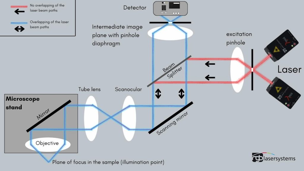
532 nm lasers are available with excellent beam quality (TEM00), low noise, high power stability, narrow linewidths and long coherence lengths, making them ideal for high-precision applications.
- Interferometry and holography: high coherence and narrow linewidth allow ultra-precise surface and dimensional measurements.
- Particle sizing and characterization: sizing particles by analyzing diffraction patterns, and evaluating the size, granularity and fluorescence of cells.
- Semiconductor wafer inspection: identifying imperfections on semiconductor wafers, measuring feature dimensions in integrated circuits, and ensuring alignment.

Measuring thermal expansion with a 532 nm Michelson interferometer
Choose a material and temperature change for a 100 mm part to see the expansion and the fringes it produces.
Less than one fringe: resolve it by phase measurement or a longer part.
Easily counted: each fringe is a 266 nm step of the mirror.
Easily counted: each fringe is a 266 nm step of the mirror.
Easily counted: each fringe is a 266 nm step of the mirror.
Less than one fringe: resolve it by phase measurement or a longer part.
Easily counted: each fringe is a 266 nm step of the mirror.
Easily counted: each fringe is a 266 nm step of the mirror.
Easily counted: each fringe is a 266 nm step of the mirror.
Less than one fringe: resolve it by phase measurement or a longer part.
Easily counted: each fringe is a 266 nm step of the mirror.
Easily counted: each fringe is a 266 nm step of the mirror.
Easily counted: each fringe is a 266 nm step of the mirror.
Less than one fringe: resolve it by phase measurement or a longer part.
Less than one fringe: resolve it by phase measurement or a longer part.
Less than one fringe: resolve it by phase measurement or a longer part.
Easily counted: each fringe is a 266 nm step of the mirror.
Less than one fringe: resolve it by phase measurement or a longer part.
Less than one fringe: resolve it by phase measurement or a longer part.
Less than one fringe: resolve it by phase measurement or a longer part.
Easily counted: each fringe is a 266 nm step of the mirror.
ΔL = α·L·ΔT; in a Michelson interferometer each fringe corresponds to a mirror displacement of λ/2 (266 nm at 532 nm). Typical room-temperature expansion coefficients.
Environmental, Remote Sensing and Satellite Applications
532 nm lasers are used in a variety of atmospheric and aquatic sensing technologies:
- Lidar: continuous or modulated green laser light is used in aerosol profiling, pollution monitoring and hydrographic mapping.
- Underwater optical communication: green wavelengths penetrate water better than red or infrared, supporting short-range data transmission and imaging in oceanographic research.
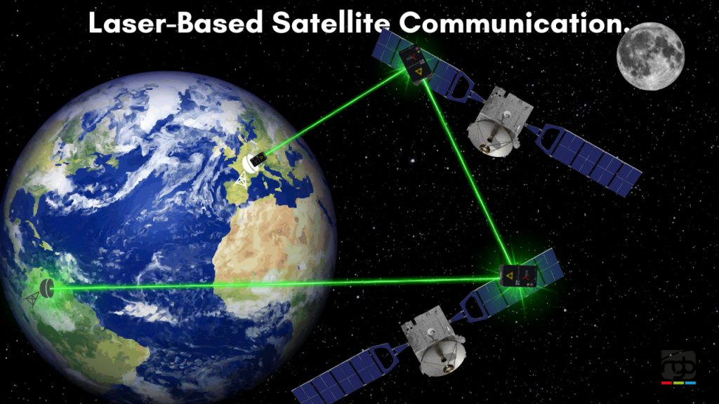
- Satellite laser ranging and communication: 532 nm is a standard wavelength for satellite laser ranging and is studied for free-space links between satellites and ground stations.
Display Technologies and Entertainment
- Laser light shows and projectors: the 532 nm beam produces brilliant green light, essential in high-visibility displays and RGB laser projection systems.
- Holographic displays and augmented reality: next-generation display technologies increasingly use compact green laser sources for sharp, vivid visuals.

Choosing a 532 nm CW Laser
Each application favors different laser properties: narrow linewidth for Raman and holography, power and stability for fluorescence, and compactness for alignment and OEM integration.
Choosing a green CW laser
Raman spectroscopy
Choose the Lambda Beam 532 nm narrow line DPSS (125, 175 or 200 mW). For fluorescent samples, consider the Lambda Beam Wavelock at 785 nm.
Fluorescence and flow cytometry
The Lambda Beam 532 nm DPSS from 75 to 200 mW excites PE, Cy3, TRITC and red fluorescent proteins; add a 488 nm Lambda Beam for FITC and GFP.
Interferometry and holography
Use a narrow line Lambda Beam 532 nm for long coherence, or a single-frequency DPSS laser from Skylark Lasers when you need sub-MHz linewidth at another wavelength.
Alignment, teaching and compact OEM
For a small, USB-powered green source, the Lambda Mini offers 520 nm diode output up to 25 mW free beam or 50 mW fiber coupled.
Choose your application.
Conclusion
From molecular fingerprinting with Raman spectroscopy to retinal therapy, microsoldering and laser displays, CW 532 nm lasers are an exceptionally versatile and powerful tool. Their blend of visibility, absorption characteristics and spectral precision continues to unlock possibilities in research, manufacturing and healthcare, and as laser technology advances, the applications of this brilliant green beam will only expand.
Contact Axiom Optics experts for more information and help choosing the right CW laser source for your application.
532 nm Laser FAQ
Why are 532 nm lasers so widely used?
532 nm combines high beam quality, a bright visible green output that is easy to align, stable power, and strong absorption by many materials, including hemoglobin and melanin. Most 532 nm CW lasers are diode-pumped solid-state (DPSS) lasers that frequency-double 1064 nm light, which gives them excellent beam quality and narrow linewidth options.
Is 532 nm a good wavelength for Raman spectroscopy?
Yes, for many samples. Raman scattering scales with the fourth power of frequency, so 532 nm gives several times more signal than 785 nm for the same power, and the Raman lines stay within the range of silicon detectors. The trade-off is a higher risk of fluorescence background in colored or biological samples, where 785 nm is often preferred.
Which fluorophores are excited at 532 nm?
532 nm efficiently excites dyes and proteins such as Rhodamine 6G, Alexa Fluor 532, propidium iodide, Cy3, TRITC, tdTomato and R-phycoerythrin (PE). Green-emitting dyes such as FITC, GFP and SYTOX Green are better excited at 488 nm.
Why does green light travel farther underwater than red or infrared?
Water absorbs red and infrared light much more strongly than blue and green light. In many natural waters the clearest window sits in the blue-green, which is why 532 nm is used for underwater optical communication and airborne bathymetric lidar.
What should I consider for 532 nm laser safety?
Use laser safety eyewear rated for 532 nm at your power level. Because most 532 nm lasers are DPSS sources, also confirm that residual 1064 nm and 808 nm infrared light is filtered or covered by your eyewear, since it is invisible.
Where can I buy a 532 nm CW laser?
Axiom Optics supplies the Lambda Beam 532 nm DPSS laser in standard and narrow line versions, along with compact diode lasers at nearby green wavelengths, and can recommend the right source for your application.
Find your 532 nm laser
Tell Axiom Optics your application, power and linewidth needs, and we will recommend the right CW laser.